MODULE 1 - INTRODUCTION
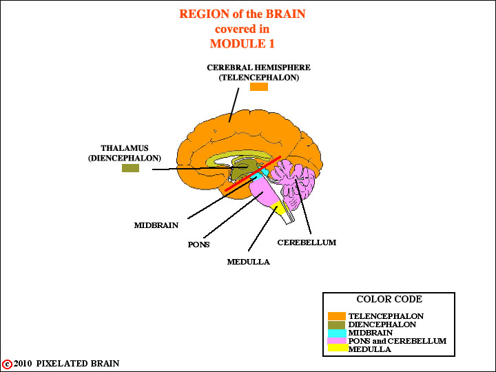
The best way to present the clinical anatomy of the brain is by taking up systems, one at a time - motor, sensory, visual, etc. But you can't trace these systems through the brain unless you have some familiarity with the "territory". For example, we will tell you, in Module 5, that the main motor pathway descends through “the pyramids, found in the ventral part of the medulla”. But what do the words pyramid, ventral and medulla really mean? So, we will use the first two modules of this program to give you a crash course in the anatomy of the brain and spinal cord.
We start Module 1 by looking at the skull and the meninges - tissue layers that protect the brain from the harsh environment of the outside world. Given the close relationship between the meninges and cerebrospinal fluid, we consider it as well. Then, we go on to the brain.
In Module 2 we will look at the diencephalon in greater detail and then go on to discuss the regions below the red line. These include the brainstem and the spinal cord.
Unfortunately, in these two modules there will be lots of names to memorize and not much time to consider brain function. For a list of them, click the "I want to" link at the bottom of this page; then click on the Glossary for Module 1.Be patient, the fun starts with Module 3 when take up somatic sensory pathways.hhh
At the bottom of this page, just below this note, is a collection of links. They are found at the bottom of most pages in the Pixelated Brain and you should experiment with them. For now, click on "Next" to continue with Module 1.
SITE MAP MOD 1 HOME PIXBRAIN HOME I WANT TO COMMENT