MODULE 1 - SECTION 1 - THE SKULL
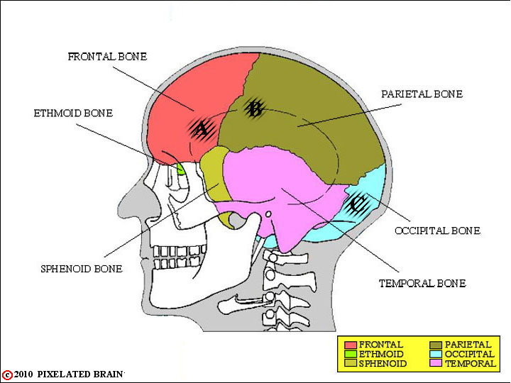
SKULL - a LATERAL VIEW
This lateral view shows the 6 bones that contribute to the formation of the cranial cavity. Of course, we really care about is the relationship of these bones to the brain. So what we show you here is three penetrating lesions of the skull - A, B and C. In the next view corresponding marks are made on the brain.
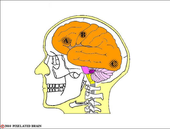
BRAIN in SKULL - a LATERAL VIEW
By the end of this module you will know that region A of the brain plays an important role in organizing
speech. So, confronted with a patient with skull lesion A, you would want to test carefully to determine if speech had been impaired as a
result.
Similarly, area B is involved motor control of the right arm, and area C is concerned with vision. Examination of each patient would focus
on testing these functions.
In short, if you can visualize the relationship between the skull and the brain you will have clues regarding what to look for when the
skull is damaged.
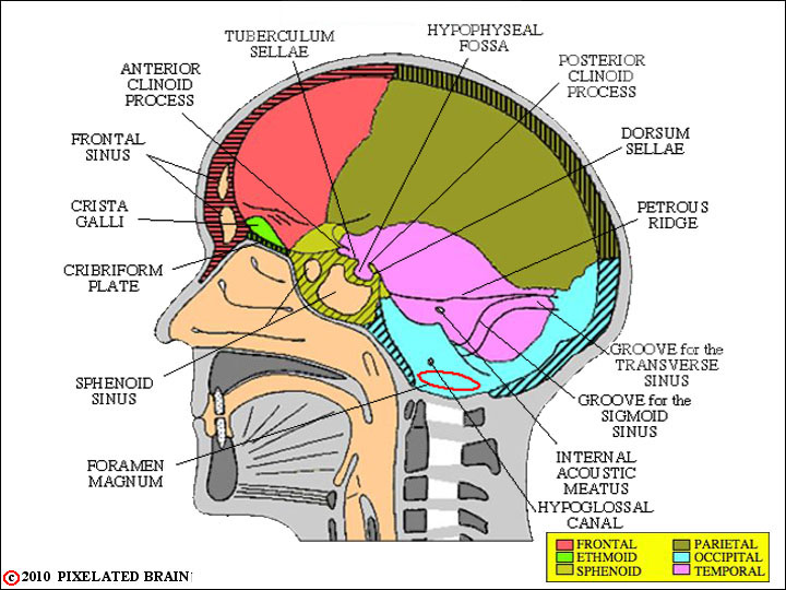
SKULL - a MIDSAGITTAL VIEW
The skull has been cut in the mid-sagittal plane, and we are within the cranial cavity, looking laterally.
Line drawings of this sort do a poor job of conveying the 3-dimensional aspects of this complex space, so if
possible compare this view with an actual skull. Note the close relationship of the frontal and sphenoid sinuses to the interior of the cranial cavity.
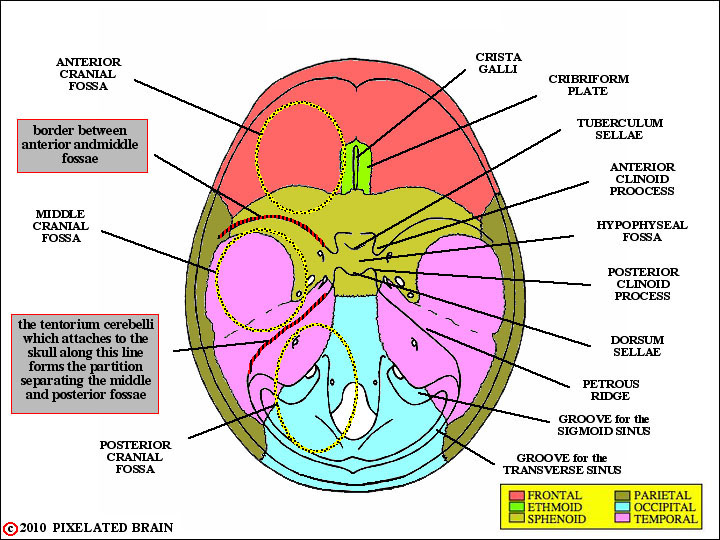
SKULL - a BASAL VIEW
In this view looks down at the floor of the cranial cavity. On the right, some of the obvious landmarks are labeled. On the left, we have tried to introduce you to the concept of the three cranial fossae. These terms are frequently used, clinically, to describe different regions within the cranial cavity.
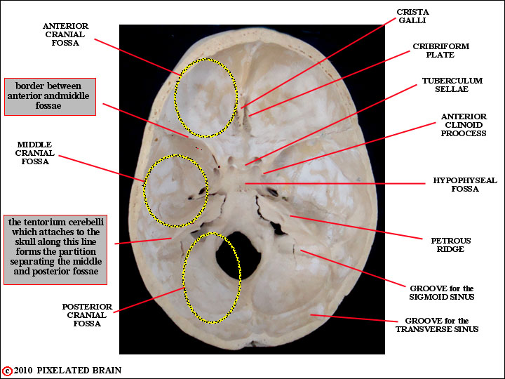
A BASAL VIEW of the HUMAN SKULL
Some landmarks in a view of the human skull.
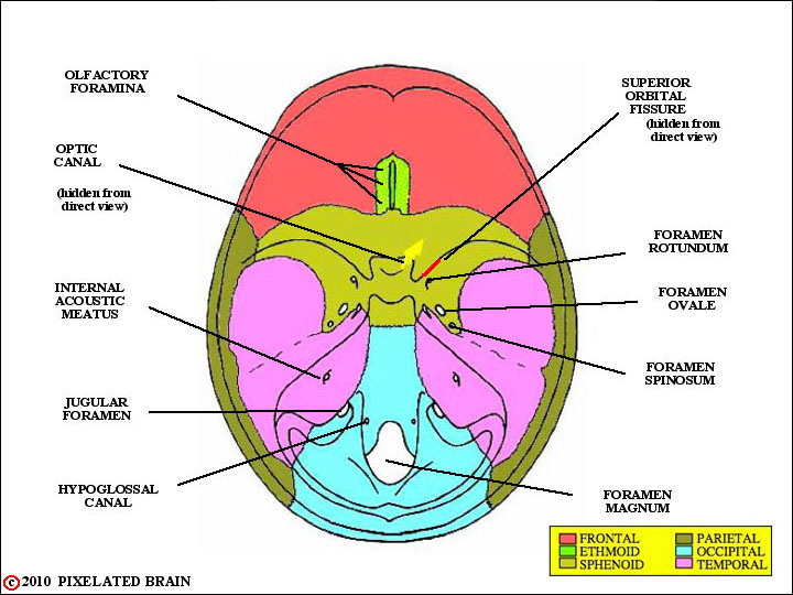
FORAMINA in the SKULL
This view simply shows the major openings in the skull. When we take up the cranial nerves, later on, you will want to be able to say which nerves or structures pass through each of these openings, but we propose to skip this detail for now.
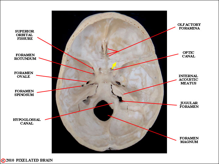
FORAMINA in the HUMAN SKULL
Here are some of the major foramina in a basal view of the human skull.