MODULE 1 - SECTION 2 - BRAIN DEVELOPMENT
The brain is actually a highly distorted tube-like structure. To understand how this comes about it is useful to take a brief look at how the brain develops.
THE NEURAL PLATE
In the first few days following fertilization of the ovum, the single celled zygote divides repeatedly to form a solid cell mass of 12 - 16 cells called a morula. During the second week of development this structure implants in the wall of the uterus. Two cavities now develop within the mass - the amnionic cavity above and the yolk sac below. Between these two lies the embryonic bilaminar disc, consisting of a layer of ectoderm lying above a layer of entoderm. During the third week of development a region termed the primitive streak forms on the dorsal surface of the disc, giving a longitudinal axis to the developing embryo. Cells from the surface of the disc migrate through this region and then spread out between the ectoderm and the entoderm to create a new cell layer. The cells that migrate laterally form mesoderm, thus transforming the bilaminar disc into a trilaminar one.
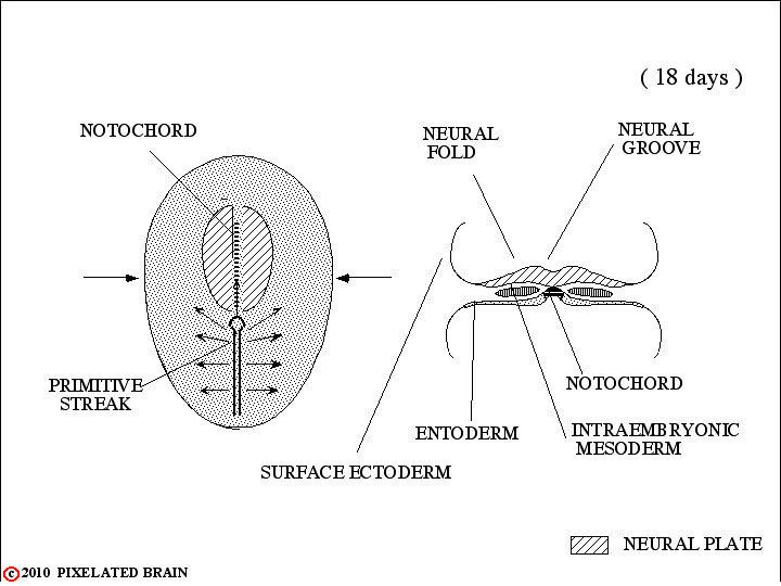
Other cells migrate anteriorly in the midline to form the notocord. By a process of neural transduction, the notocord induces the overlying ectoderm to become neural tissue - the neural plate. In the midline, just above the notocord, a neural groove forms. Lateral to this on either there is a proliferation of neural ectoderm to form neural folds.
THE NEURAL TUBE
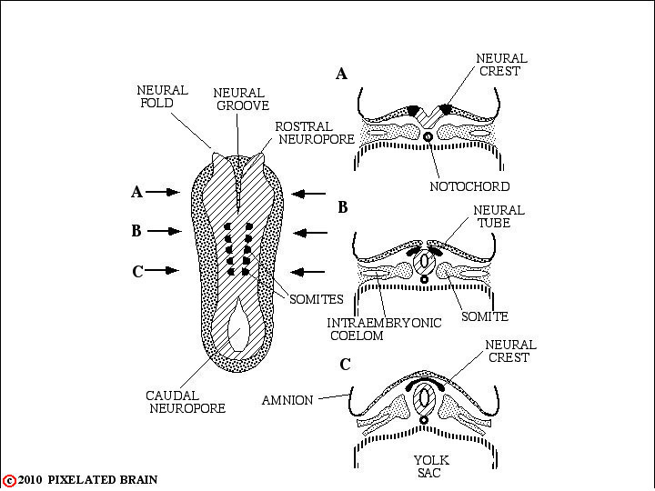
On about day 18 following fertilization, the process of neurulation begins. Starting in the cervical region, the neural folds migrate dorsally and fold toward the midline to meet and fuse, forming the first part of the neural tube. Fusion of the folds then extends both rostrally and caudally. For several days the ends of the neural tube remain open.The rostral opening, or cranial neuropore, is closed at about day 25 by a membrane called the lamina terminalis. The caudal neuropore closes a few days later.
Failure of the cranial neuropore to close leads to anencephaly, a developmental defect which grossly distorts the development of the brain. Failure of the caudal neuropore to close is associated with some form of spina bifida.
Some tissue of the neural plate fails to become encorporated in the neural tube and remains just dorsolateral to it. This neural crest tissue ultimately gives rise to many structures, including: 1) the dorsal root (sensory) ganglia of the peripheral nervous system; 2) the ganglia of the autonomic nervous system; 3) the cells of the adrenal medulla and 4) the Schwann Cells that form the myelin that surrounds peripheral nerves. Lateral to the neural tube the mesoderm becomes organized in a segmental fashion into somites which will, ultimately, impose a segmental pattern upon the peripheral nervous system.
THE THREE PRIMARY VESICLES
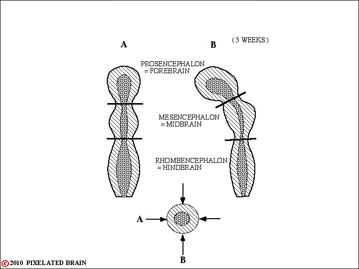
The rostral end of the neural tube undergoes a series of regional expansions to generate the final form of the brain. In early development (3-4 weeks) there are three primary vesicles:
1 - Forebrain = Prosencephalon
2 - Midbrain = Mesencephalon
3 - Hindbrain = Rhombencephalon
THE FIVE SECONDARY VESICLES
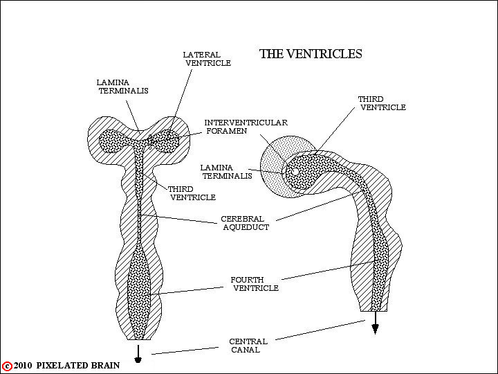
By the the fifth week of development the lateral wall of the prosencephalon has pushed outward in a blister-like manner, carrying the cental cavity with it to form a new space called the lateral ventricle. The part of the cavity that remains behind, in the mid-line, becomes the third ventricle. There is also a less than obvious subdivision of the space within the rhombencephalon, forming the rostral and caudal parts of the fourth ventricle.
Thus, we now have 5 cavities within the brain:
1 - The lateral ventricle
2 - The third ventricle
3 - The cerebral aqueduct
4 - The rostral fourth ventricle
5 - The caudal fourth ventricle.
These spaces will form the ventricular system of the brain, and as we shall see in the next view, the tissue that is the wall of each space becomes a major subdivision of the adult brain. Rostrally (i.e., at the very "top"of the neural tube) the lamina terminalis seals the ventricular system closed.
SUBDIVISIONS of BRAIN
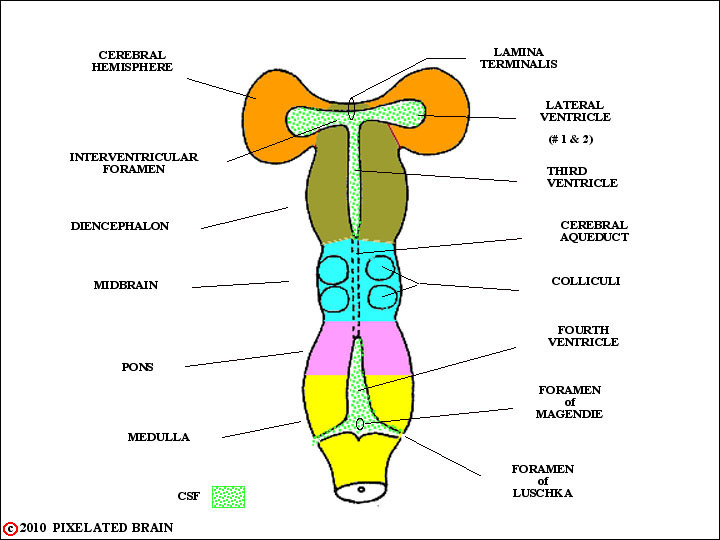
The neural tissue surrounding the developing ventricular system proliferates to become the major subdivisions of the adult brain. These regions, labeled on the left in this figure, will become the major subdivisions of the adult brain.
Note that the lateral ventricles communicate with the third ventricle by means of a constricted channel called the interventricular foramen.
Also note that the interior of the neural tube (the ventricular system) has openings to the "outside" at the level of the medulla. They are a single opening in the midline - the Foramen of Magendie - and paired lateral openings - the Foramina of Luschka.
The CHOROID PLEXUS
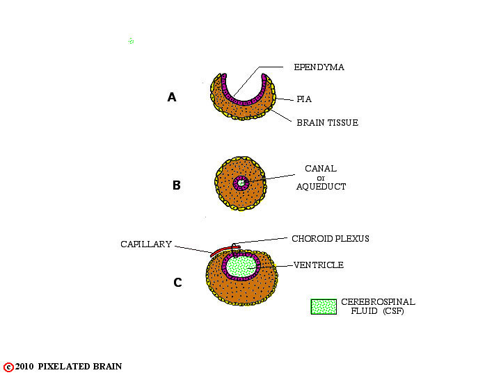
A shows us, once again, an early stage in the formation of the neural tube. At the spinal cord level, the folds meet and neural tissue completely surrounds a small, centrally placed cavity, called the spinal canal - as seen in B.
At the rostral end of the neural tube - the future brain - the story is more complex as shown in the figure just above. In one region, the midbrain, the edges of the folds meet, just as they do in the spinal cord; the small cavity within is called the cerebral aqueduct.
In other regions the edges of the neural plate fail to meet, even in the normal brain. Here, the ependymal cell layer, lining the cavity, and the layer of pial cells, covering the surface of the tube, meet to form a membrane which completes closure of the cavity, as seen in C. Blood vessels extend into this membrane creating highly vascular tissue called choroid plexus.
The choroid plexus is the tissue responsible for the formation of cerebrospinal fluid; this fluid (CSF) fills the ventricular system and escapes to the "outside" by passing through the Foramina of Luschka and Magendie.
CHOROID PLEXUS "ROOFS"
THREE VENTRICLES
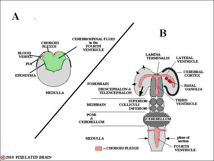
A is a slightly more realistic cross-section through the medulla; the plane of section is shown in B. At this level blood vessels invade the layer formed by the fusion of the pia and the ependyma to create the choroid plexus.
B is a dorsal view of the developing brain, similar to Fig.1.2.5, but providing added detail for each region:
- Medulla - choroid plexus forms the roof of the third ventricle.
- Pons - the neural folds fail to meet (not shown) but rather give rise to a new structure, the cerebellum. The greatly expanding Cerebellum lies in the midline, dorsal to the rostral extension of the Fourth Ventricle. Together with pathways that connect it to the rest of the brain - the cerebellar peduncles - it, rather than choroid plexus, forms the roof of the ventricle.
- Midbrain - neural tissue enclose a small passageway, the cerebral aqueduct.
- Diencephalon and Telencephalon - choroid plexus roofs of the third ventricle and is carried out into the lateral ventricles within the hemisphere..
THE BRAIN "BENDS"
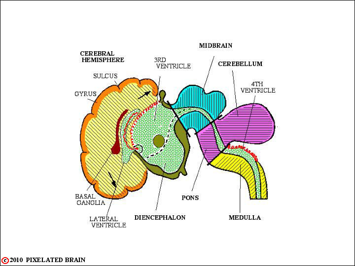
This is a side view of the brain, at about the same stage of development as the one above. Things to note are:
-The choroid plexus does indeed form the roof of the fourth and third ventricles.
-It can be seen passing out through the interventricular foramen (a small black circle, not labeled) into the lateral ventricle.
-The brain now makes a sharp bend at the midbrain level - a process that started much earlier.
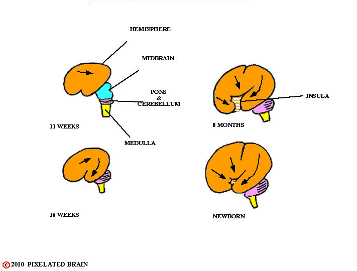
-The cerebral hemisphere is now greatly expanding, as shown by the arrows, and it will eventually cover the brainstem, hiding it from view.
-As the hemisphere enlarges, the surface becomes "wrinkled" with many convex gyri separated from each other by depressions called sulci.

EXPANDING HEMISPHERE
"BURIES"
THE INSULA
With time, the future frontal and temporal lobes migrate (red arrow) and meet. The narrow gap between them becomes the lateral fissure and in the depth of the fissure lies a buried part of the hemisphere's surface called the insula.
FRONTAL and TEMPORAL LOBES
MEET
CREATING the LATERAL FISSURE
The insula is now almost completely hidden from view.