MODULE 1 - SECTION 3 - THE MENINGES
1
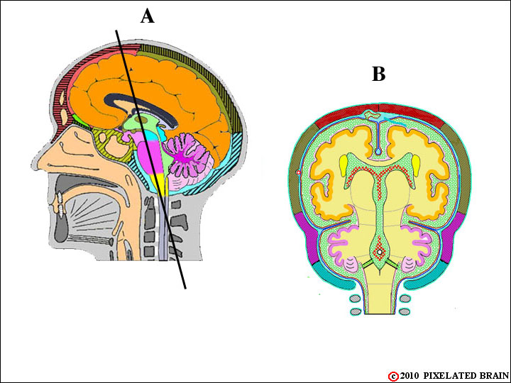
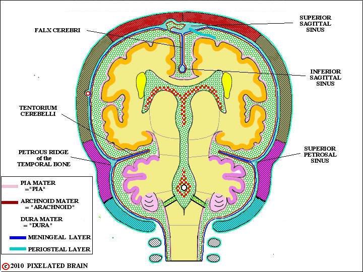
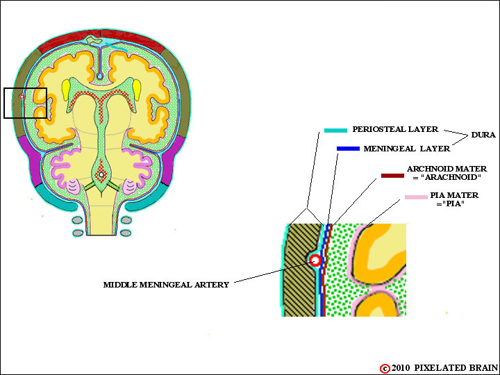
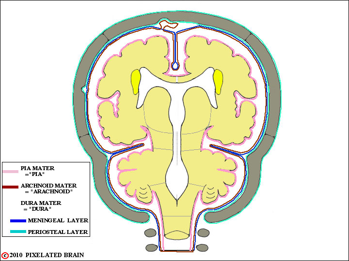
THE BASIC FRONTAL SECTION - I
Certainly, the most important function of the skull is to protect the fragile brain, lying within the cranial cavity. As this view demonstrates, the skull is well-designed for the job. If we cut through the skull in the plane shown in A and look at the surface of the cut we get view B. The contour of the brain's surface matches that of the cranial cavity rather well, but there is a small space between the two. Within this space lie three membranes known collectively as the meninges; they include the dura mater (aka dura), the arachnoid mater (aka arachnoid) and the pia mater (aka pia). Filling the remaining space is a clear colorless liquid called cerebrospinal fluid (CSF). The brain is, in a sense, floating in CSF which protects it from "bumping into" the skull during sudden movements of the head. From a clinical point of view, the anatomy of this region is extremely important and we want to look at it in detail.
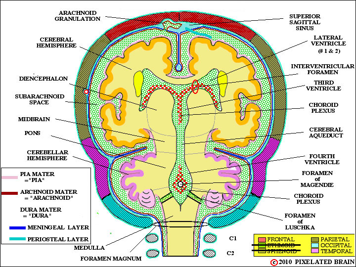
THE BASIC FRONTAL SECTION - II
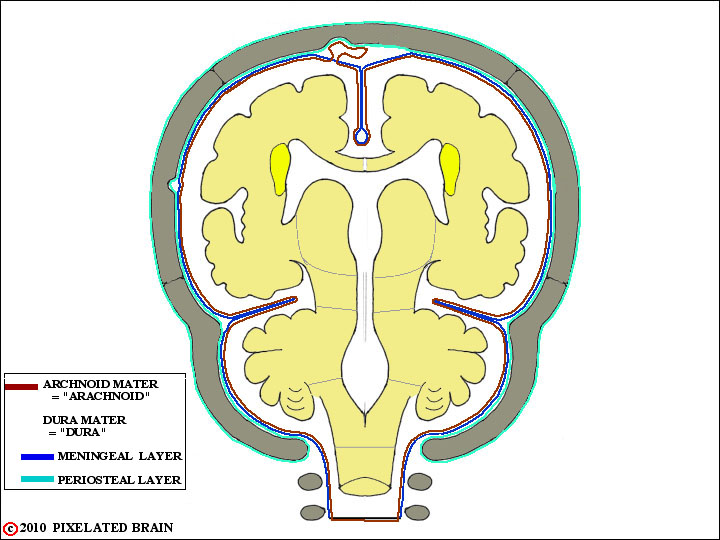
Here's an enlarged version of our frontal section. It is a "summary view", crowded with labels and detail. Don't spend too much time on it now, but come back to it from time to time as our discussion progresses.
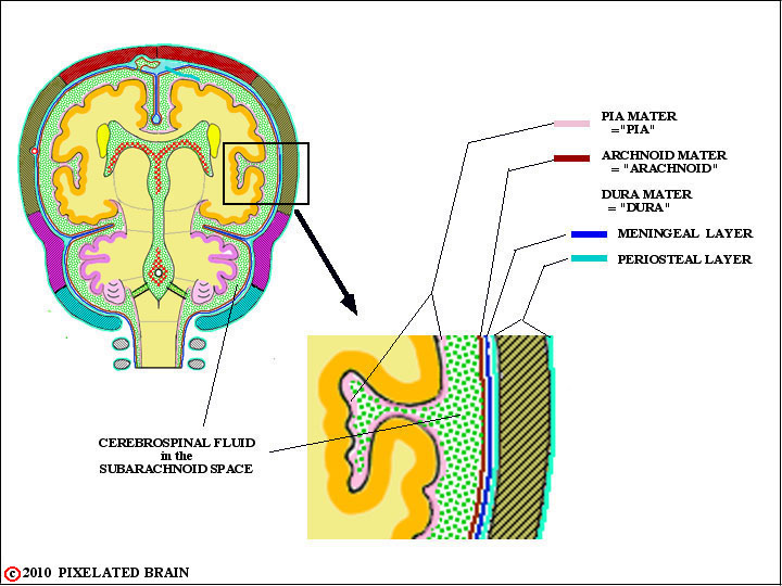
THE MENINGES
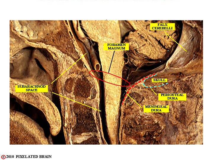
The outermost meningeal layer is the dura. It is composed of dense fibrous connective and has the texture and strength of canvas. It consists of an outer, periosteal layer and an inner, meningeal one. In most regions the two layers are fused together, and appear to be a single one.
Within the skull, the dura is tightly adherent to the overlying bone, and this attachment is particularly strong in regions where the membrane crosses a bony suture line.
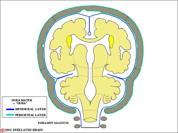
ANOTHER VIEW OF THE PERIOSTEAL AND MENINGEAL LAYERS OF DURA
THE DURA MATER
In some regions the meningeal layer of the dura breaks away from the dural one to form tough, unyielding folds or partitions which partially subdivide the cranial cavity. Two of these - the falx cerebri and the tentorium cerebelli- are shown here. These partitions are important because when expanding lesions in the cranial cavity - blood or a tumor - displace brain tissue, it is often obliged to herniate around one of them. For a look at the falx cerebri and tentorium cerebelli in a dissected specimen, see DiganatA_1.13 .
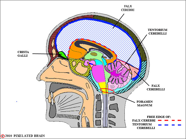
FALX CEREBRI
and
TENTORIUM CEREBELLI
THE VENOUS SINUSES
When the dural layers separate, as described above, they often enclose a network of venous sinuses. Here the superior sagittal sinus, the inferior sagittal sinus and the superior petrosal sinus are labeled..
THE VENOUS SINUSES
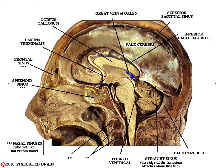
Another example, in which the meningeal dura encloses a sinus, is the straight sinus. In this case, the sinus is created in the region where the falx cerebri joins the ridge line of the tentorium cerebelli. See Haines, pgs.110-112 for more diagrams and details.
MIDDLE MENINGEAL ARTERY
The layers of the dura also split to enclose the middle meningeal artery. The split is asymmetrical in that the meningeal surface - the one that the brain rubs against when it moves slightly within the cranial cavity - remains smooth, but the vessel creates a ridge in the periostial surface.
The result of this asymmetrical split is that the artery erodes a "valley" in the inner surface of the skull. If the skull fracture and the fracture line crosses this valley, then there is a good chance the middle meningeal artery will be torn and that arterial blood will leak forming a hematoma (pool of blood) between the dura and the skull.
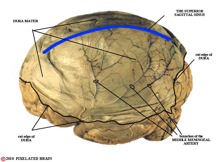
DURA MATER
showing course of
MIDDLE MENINGEAL ARTERY
To prepare this specimen, the brain was carefully removed from the skull; then a large piece of dura was detached from the skull and placed on the brain.
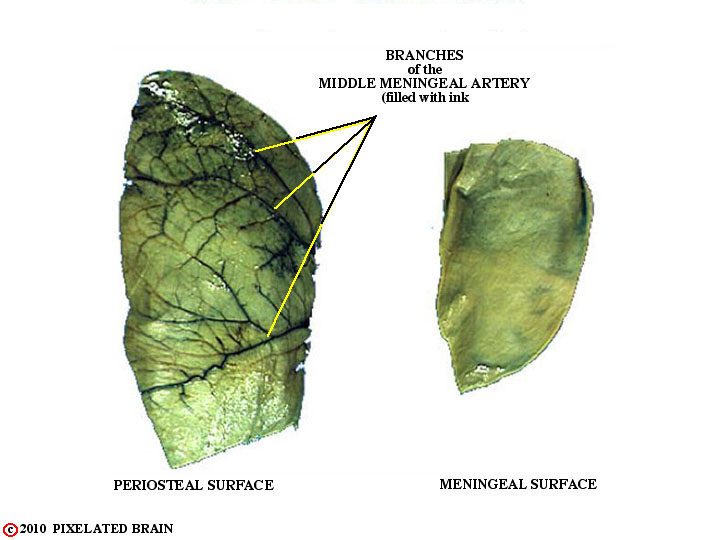
THE TWO SURFACES
of the
DURA MATER
The dura, shown in the specimen above, was subsequently removed from the surface of the brain and cut into these two pieces. Then india ink was injected into the middle meningeal artery. Note how the branches of the vessel stand out on the outer ( = periosteal ) surface. If you rubbed your finger over it, you would easily feel these ridges. In contrast, the inner ( = meningeal ) surface is quite smooth. When the brain moves slightly within the skull (when you bump your head against a wall, for example) the brain's surface moves, relative to the inner surface of the dura, and it makes sense for the latter to be smooth. The result of this asymmetrical split is that the artery erodes a "valley" in the inner surface of the skull. If the skull fracture and the fracture line crosses this valley, then there is a good chance the middle meningeal artery will be torn and that arterial blood will leak forming a hematoma (pool of blood) between the dura and the skull.
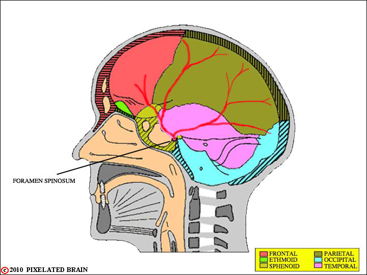
INTERIOR of the SKULL
showing course of the
MIDDLE MENINGEAL ARTERY
The middle meningeal artery enters the the cranial cavity through the foramen spinosum. For a better view of the foramen, click here.
THE SPINAL DURA is DIFFERENT
The
THE DURA MATER
is
PAIN SENSATIVE
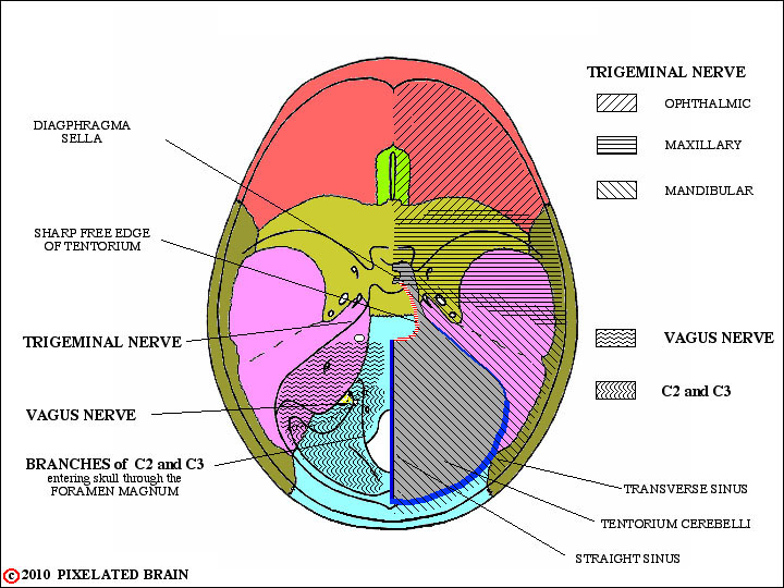
The dura is a pain-sensitive structure - but the arachnoid, pia and the brain itself are not. The dura of the anterior and middle fossae receives sensory endings from the trigeminal nerve. The dura of posterior cranial fossa is supplied by branches of the vagus nerve, coming off before it exits from the cranial cavity) and by branches of spinal nerves C2 and C3, that enter through the Foramen Magnum. The significance of this is that irritation of the dura may be perceived (or "referred" to) as coming from sensory territory of the trigeminal, vagal or upper cervical nerves.
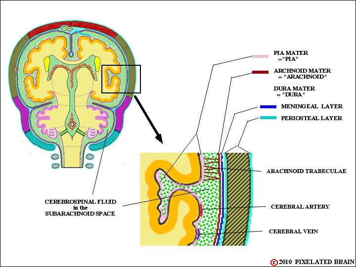
THE ARACHNOID view 1
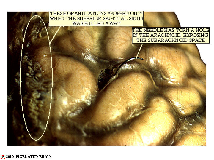
The arachnoid is a delicate membrane that lies just deep to the dura. It is a fluid- tight structure, and - in life - is pushed against the dura by the pressure of the cerebrospinal fluid. A network of fine collagenous fibers, resembling a spider web, pass between the arachnoid and the pia, giving this layer its name (arachnoid = Greek for spider). Except for the region close to the superior sagittal sinus, there are no structures connecting the arachnoid with the dura. Thus
THE ARACHNOID view 2
THE ARACHNOID view 3
THE PIA
The
THE PIA
16