MODULE 1 - SECTION 6 - INTERNAL ANATOMY of the CEREBRAL HEMISPHERE

One of the most difficult topics for students of neuroanatomy to master has to do with the internal anatomy of the cerebral hemispheres. There are actually only a few structures (and names) involved, but the manner in which the brain has developed has caused the relationships between them to be complex. Added to this is the fact that the most convenient way to study the hemisphere involves the use of serial sections - and that no matter what plane of section one picks, the cuts will pass through some structures at odd angles. The end result for the student is confusion which usually does not begin to lift until the last weeks of the course. We hope that by focusing on this subject at the start we will help you avoid this. But, be prepared for some tough going!

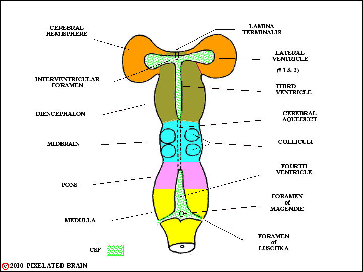
     Early in the module we felt obliged to say a little about brain development because we wanted to explain how the ventricular system came to be. So, when we last looked at the developing hemisphere, in this figure, it was just a blister, extending laterally from the diencephalon. In fact, things are more complicated, as shown next, in a schematic frontal section through the hemisphere and diencephalon

1
     A sheet of cells, the cerebral cortex, lies just below the cortical surface. Underneath the cortex lies the "white matter" - actually a jumble of axon bundles running every which-way. Buried within the white matter are several nuclei - each of them a region of densely packed neuron cell bodies. As the axons of the white matter try to pass to and from the cortex, and from one cortical region or hemisphere to another, they must pass between and around these so-called deep nuclei. The nuclei are, in a sense, the troublemakers; as a group, they are known as the basal ganglia and they include the caudate nucleus, the putamen, the globus pallidus, the amygdala and the claustrum. The major white matter pathways threading between the nuclei include:
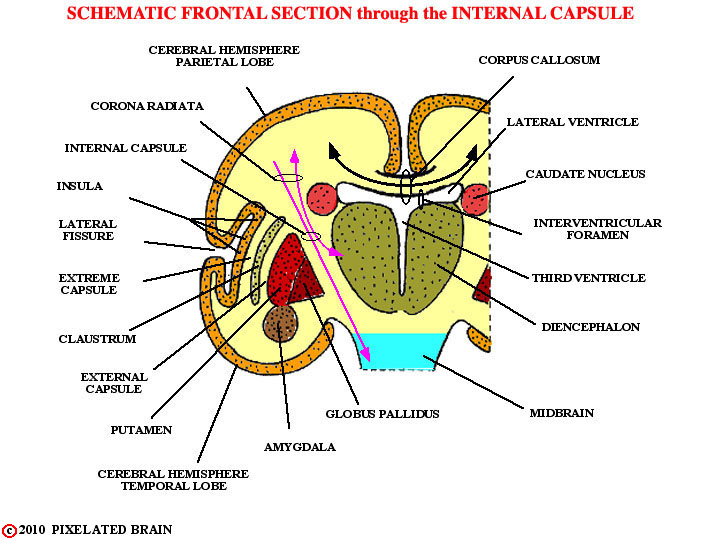
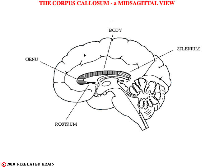
   1) the corpus callosum, connecting one cerebral hemisphere with the other and often forming the roof of the lateral ventricle.
    2) the internal capsule, which contains some fibers (axons) interconnecting the diencephalon with the cerebral cortex and others which pass from the cortex to more caudal parts of the brainstem and the spinal cord.
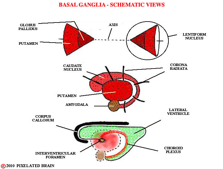
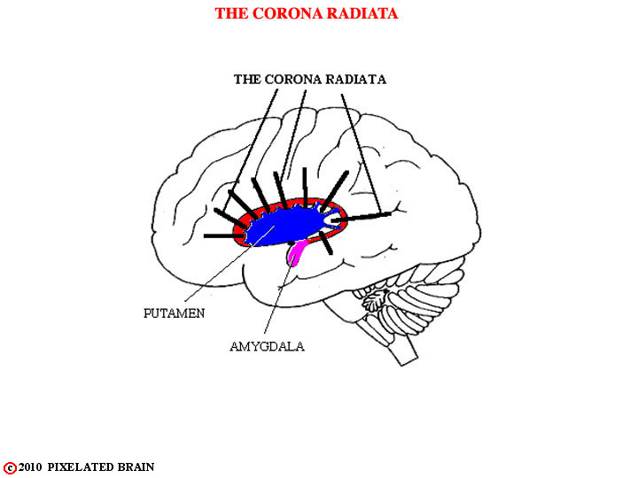
    3) the corona radiata, which represents the extension of the internal capsule toward the surface of the hemisphere, into the region where the fibers are no longer trapped between the caudate and the lentiform nucleus.
    4) the external and extreme capsules, pathways connecting one part of the hemisphere with another and passing on either side of the claustrum.
2
 
3
 
4
 
5
 
6
 
7
 
8
 
9
 
10
 
11
 
12
  Whole Brain Sections in the Frontal Plane wholebrfr.htm
13
  Whole Brain Sections in the Horizontal Plane wholebrhor.htm
14
                  FIGURE 1.54
     The
15
                  FIGURE 1.55
     The
16
                  FIGURE 1.56
     The
17
                  FIGURE 1.57
     The
18
                  FIGURE 1.58
     The
19
                  FIGURE 1.59
     The
20
                  FIGURE 1.60
     The
21
                  FIGURE 1.61
     The
22
                  FIGURE 1.62
     The
23
                  FIGURE 1.63
     The
24
                  FIGURE 1.64
     The
25
                  FIGURE 1.65
     The
26
                  FIGURE 1.66
     
Clearly
27
                  FIGURE 1.41
    
 The
28
                  FIGURE 1.42
     This
29
                  FIGURE 1.43
    The
30
                  FIGURE 1.44
     The
31
                  FIGURE 1.45
     The
32
                  FIGURE 1.46
     The
33
                  FIGURE 1.47
     The
34
                  FIGURE 1.48
     The
35
                  FIGURE 1.49
     The
36
                  FIGURE 1.50
     The
37
                  FIGURE 1.51
     The
38
                  FIGURE 1.52
     The
39
                  FIGURE 1.53
     The
40
                  FIGURE 1.54
     The
41
                  FIGURE 1.55
     The
42
                  FIGURE 1.56
     The
43
                  FIGURE 1.57
     The
44
                  FIGURE 1.58
     The
45
                  FIGURE 1.59
     The
46
                  FIGURE 1.60
     The
47
                  FIGURE 1.61
     The
48
                  FIGURE 1.62
     The
49
                  FIGURE 1.63
     The
50
                  FIGURE 1.64
     The
51
                  FIGURE 1.65
     The
52
                  FIGURE 1.66
     
Clearly
53

SITE MAP      MOD 1 HOME      PIXBRAIN HOME       I WANT TO

NEXT

1 2 3 4 5 6 7 8 9 10 11 12 13 14 15 16 17 18 19 20 21 22 23 24 25 26 27