MODULE 2 - SECTION 1 - THE BRAINSTEM
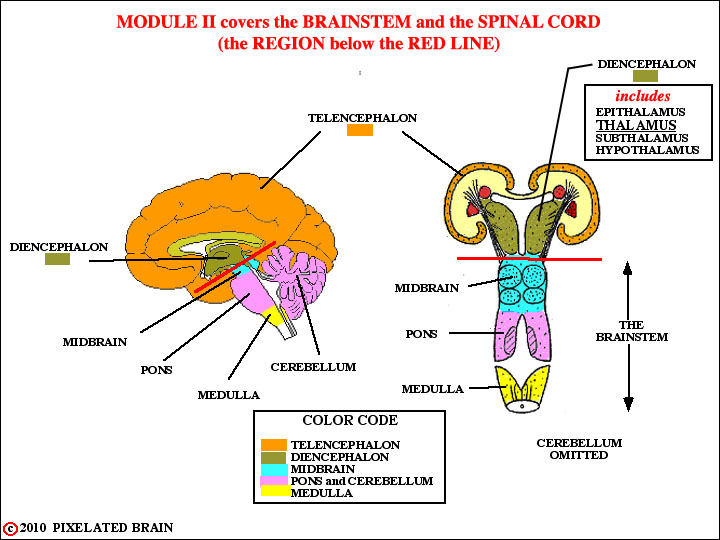
Probably we should start by defining the brainstem. This first figure shows that it consists of all the brain caudal to the forebrain (excluding the cerebellum). Practically, however, the thalamus (part of the Diencephalon) is so closely related to the brainstem that we are going to include it in our discussion.
The brain is, from an embryological point of view, the rostral end of the neural tube - continuous caudally with the spinal cord in the region of the foramen magnum. Unlike the spinal cord, in which each segment looks more or less like the next one, regional differences in development give each division of the brainstem its own peculiar appearance.
Note that the color code we will use to identify regions of the brain in most of our drawings is given here.
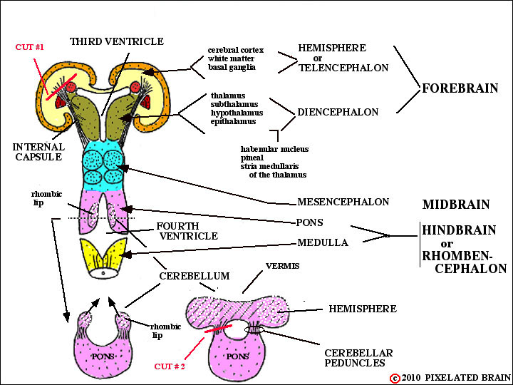
You are already familiar with the way in which the cerebral hemisphere develops by evaginating outward from the side wall of the diencephalon.
The cerebellum, in contrast, develops as a proliferation of the cells in the edge of the neural fold (the so-called rhombic lip, because it forms a margin of the rhomboid - shaped fourth ventricle) at the pontine level. The cell masses on either side join in the midline to become the cerebellum. Unlike the cerebral hemisphere, the cerebellum has a midline component (the vermis) as well as two lateral extensions, the cerebellar hemispheres. Both the cerebral hemisphere and the cerebellum are "anchored" to the brainstem by massive bundles of fibers (axons) which are, in fact, the input and output pathways of these structures. In the case of the cerebral hemisphere, this fiber bundle is the internal capsule, which you have already studied. The cerebellum has 3 separate fiber bundles of this sort, known as the cerebellar peduncles.
In any attempt to look at the dorsal and lateral surfaces of the brainstem, the first problem to be confronted is that this region is hidden below the cerebral hemispheres and cerebellum. You will recall that the expanding cerebral hemisphere extends caudally to completely cover the diencephalon and midbrain. How can we expose the brainstem to view?
The obvious answer is to dissect away the tissue of the cerebral hemisphere (and of the cerebellar hemisphere, as well) which gets in the way. The most "pure" approach , at least in conceptual terms, would be to cut the relevant fiber bundles and then just lift off the hemispheres (cerebral and cerebellar). A careful look at this figure shows where to make the cuts. After making cut #1 (and this is pretty schematic) you would be able to lift off the cerebral hemisphere, leaving behind the basal ganglia, the diencephalon and the brainstem. After making cut #2 you would be able to lift off the cerebellum.
The result would be to give you a good exposure of the dorsal surface of the brainstem (plus the basal ganglia and diencephalon). In fact, this is exactly what we propose to do.
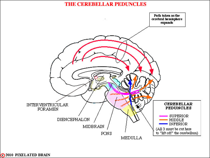
This view shows:
1) how the expansion of the cerebral hemisphere has covered the brainstem (and cerebellum), and
2) how the cerebellum is anchored to the brainstem by the three cerebellar peduncles.
Look carefully and you will see where the cut must be made to lift off the cerebellum.
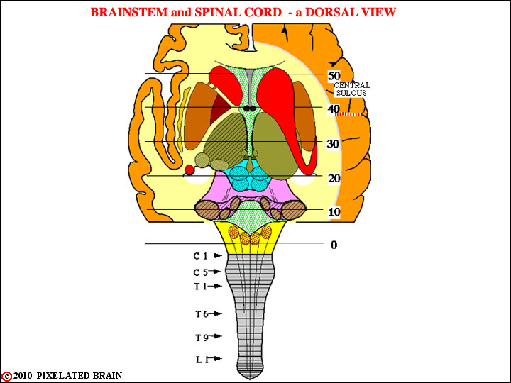
In this view, the cuts have been made and both the cerebral hemispheres and the cerebellum have been lifted off.
We see the brainstem (same color code as above) and also get another look at the basal ganglia, buried within the cerebral hemisphere.
As we explore the structure of the brain and spinal cord in future modules we will rely heavily on cross sections through these regions. The lines, numbered 0-50, define the level of the serial sections through the brain.
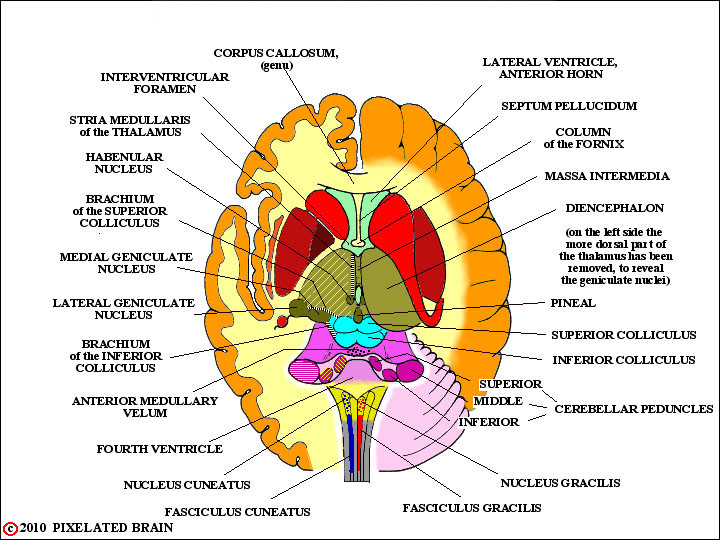
Here's a similar view in which the spinal cord has been removed and a great many labels added. Note that the individual cerebellar peduncles (cut) are identified.
You are about to be confronted with the names for a great many structures, without being told much about the function of these structures. For the moment we have two goals:
1) we want you to begin to relate a name to a structure, so that when we do take up functional issues you will at least have an idea of where the structures are in the brainstem, and
2) we want you to begin making the transition from surface view of the brainstem to cross sections through the same region.