MODULE 6 - SECTION 1 - GROSS FEATURES of the CEREBELLUM
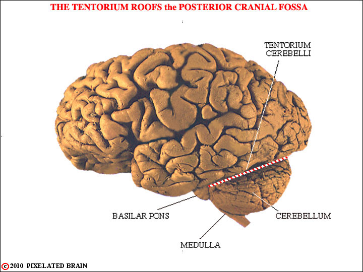
Let's begin by looking at a lateral view of the brain that you have seen before.
This should simply remind you of where the cerebellum is and shows that while it appears to lie in direct contact with the occipital lobe of the cerebral hemisphere, the two are actually separated by the tough, rigid tentorium cerebelli. The cerebellum occupies most of the space in the posterior cranial fossa. It lies dorsal to the brainstem and is connected to it by three cerebellar peduncles. It forms a roof for much of the fourth ventricle.
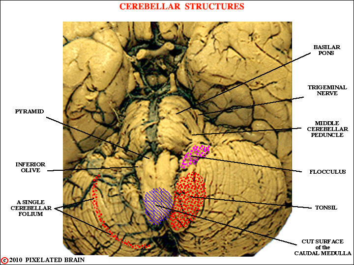
This view is of the basal surface of the brain.
Note the long extent of a folium, the position of the flocculus and the tonsil, and the fact that the trigeminal nerve exits through the middle cerebellar peduncle.
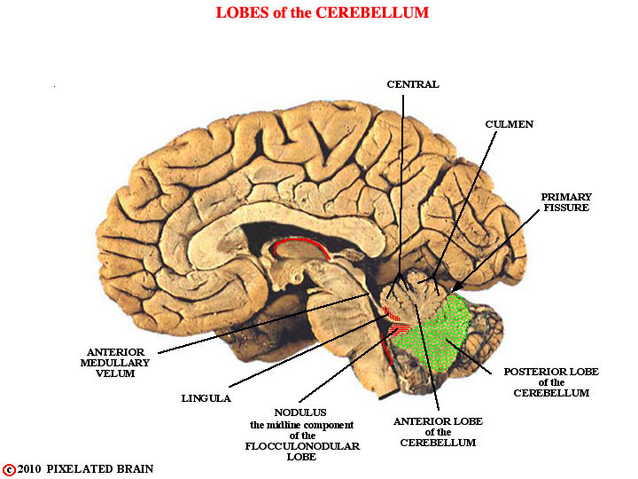
Here, we see a mid-sagittal view of the brain.
Observe that while the lingula and the nodulus are not far apart, they become widely separated when the cerebellum is "unrolled", or flattened out, as it is in many text figures.
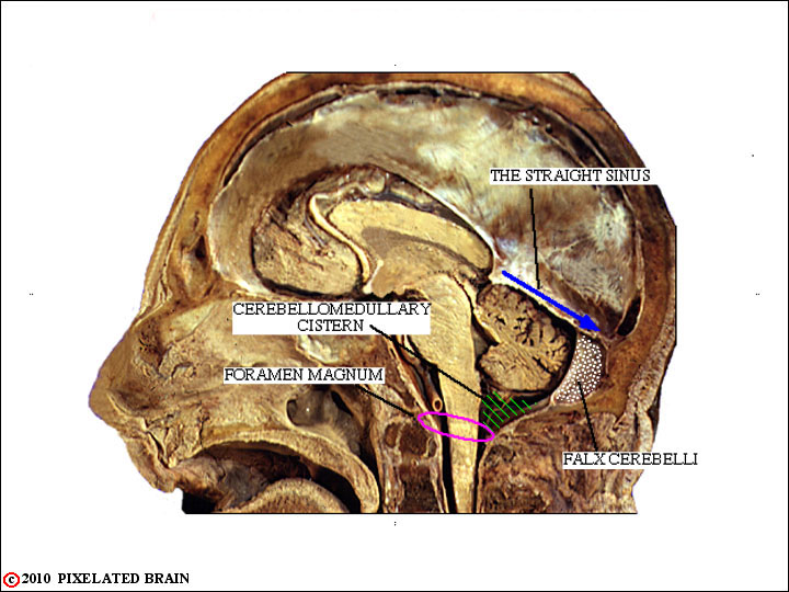
Here we see the cerebellum within the skull.
Note that the straight sinus lies just above the cerebellum, the falx cerebelli partially separates the two posterior lobes, and that a cistern of CSF and the foramen magnum lie just below.

We include this frame for two reasons.
The first is to point out that the tonsil of the cerebellum is situated just above the foramen magnum. The situation depicted here does occur and it is a true medical emergency.
The second reason for including this frame is to make the transition to sections through the cerebellum. The dotted line here shows the orientation of the slides we will be working with.
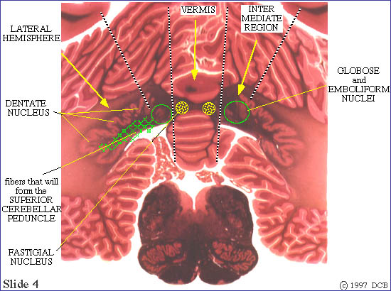
A quick look at this section tells us something about the internal anatomy of the cerebellum.
The cerebellum is divided into lateral, intermediate and vermal zones.
Each projects to its own set of deep nuclei, and the function carried out by each zone is distinct from that of the others.
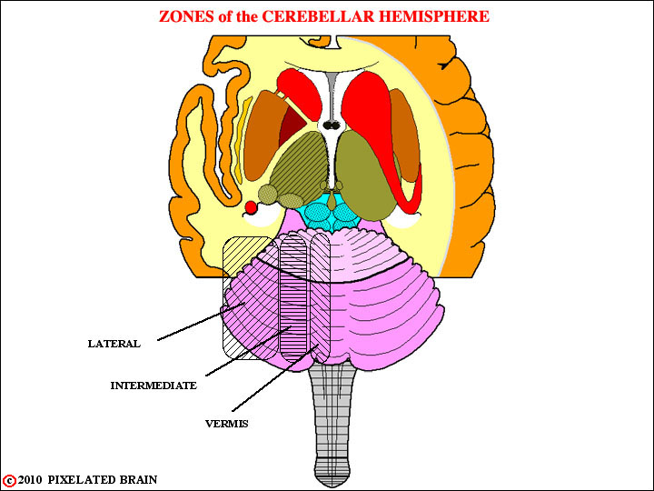
Here, we see these divisions, drawn on a surface view of the cerebellum.
1. The lateral hemisphere or cerebrocerebellum, which receives its input from the contralateral cerebral hemisphere, projects back to this same structure and is involved in motor planning.
2. The intermediate (paramedian) region or spinocerebellum which receives inputs both from the contralateral cerebral hemisphere and the ipsilateral spinal cord. This region is concerned with the adjustment of motor activity involving the distal parts of the body.
3. The midline region or vermis. This region actually carries out two rather different functions. It shares with the intermediate region the job of adjusting motor activity, in this case involving proximal and axial muscles. Parts of the vermis, however, have reciprocal connections with the vestibular nuclei. This subdivision of the vermis is termed the vestibulocerebellum and is concerned with monitoring and adjusting vestibular reflexes.
Each region of the cerebellar cortex projects out of the cerebellum by way of its own "deep" nucleus.