MODULE 6 - VIEWS-ALL
The
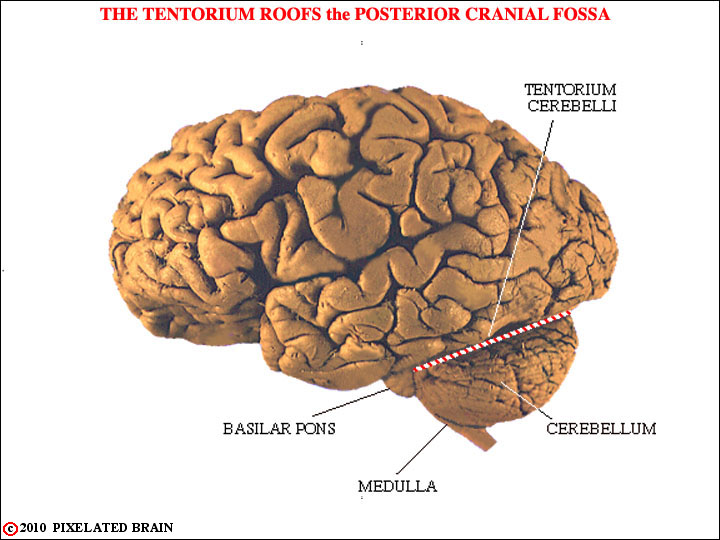
FIGURE 6-1
This simply reminds you of where the cerebellum is and shows that while it appears to lie in direct contact with the occipital lobe of the cerebral hemisphere, the two are actually separated by the tough, rigid tentorium cerebelli. The cerebellum occupies most of the space in the posterior cranial fossa. It lies dorsal to the brainstem and is connected to it by three cerebellar peduncles. It forms a roof for much of the fourth ventricle.

FIGURE 6-2
Note the long extent of a folium, the position of the flocculus and the tonsil, and the fact that the trigeminal nerve exits through the middle cerebellar peduncle.

FIGURE 6-3
Observe that while the lingula and the nodulus are not far apart, they become widely separated when the cerebellum is "unrolled", or flattened out, as it is in many text figures.
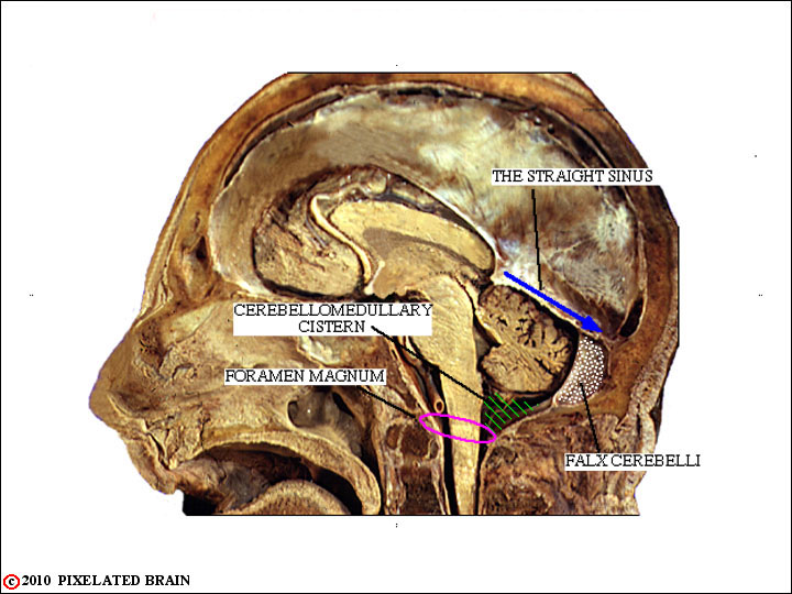
FIGURE 6-4
Note that the straight sinus lies just above the cerebellum, the falx cerebelli partially separates the two posterior lobes, and that a cistern of CSF and the foramen magnum lie just below

FIGURE 6-5
We include this frame for two reasons. The first is to point out that the tonsil of the cerebellum is situated just above the foramen magnum. The situation depicted here does occur and it is a true medical emergency. The second reason for including this frame is to make the transition to sections through the cerebellum. The dotted line here shows the orientation of the slides we will be working with.
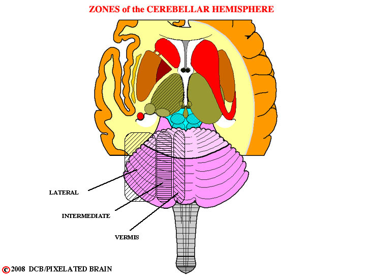
FIGURE 6-6
The cerebellum is divided into lateral, intermediate and vermal zones. Each projects to its own set of deep nuclei, and the function carried out by each zone is distinct from that of the others. This view shows these divisions,drawn on a surface view of the cerebellum.
1. The lateral hemisphere or cerebrocerebellum, which receives its input from the contralateral cerebral hemisphere, projects back to this same structure and is involved in motor planning.
2. The intermediate (paramedian) region or spinocerebellum which receives inputs both from the contralateral cerebral hemisphere and the ipsilateral spinal cord. This region is concerned with the adjustment of motor activity involving the distal parts of the body
3. The midline region or vermis. This region actually carries out two rather different functions. It shares with the intermediate region the job of adjusting motor activity, in this case involving proximal and axial muscles. Parts of the vermis, however, have reciprocal connections with the vestibular nuclei. This subdivision of the vermis is termed the vestibulocerebellum and is concerned with monitoring and adjusting vestibular reflexes.
Each region of the cerebellar cortex projects out of the cerebellum by way of its own "deep" nucleus.
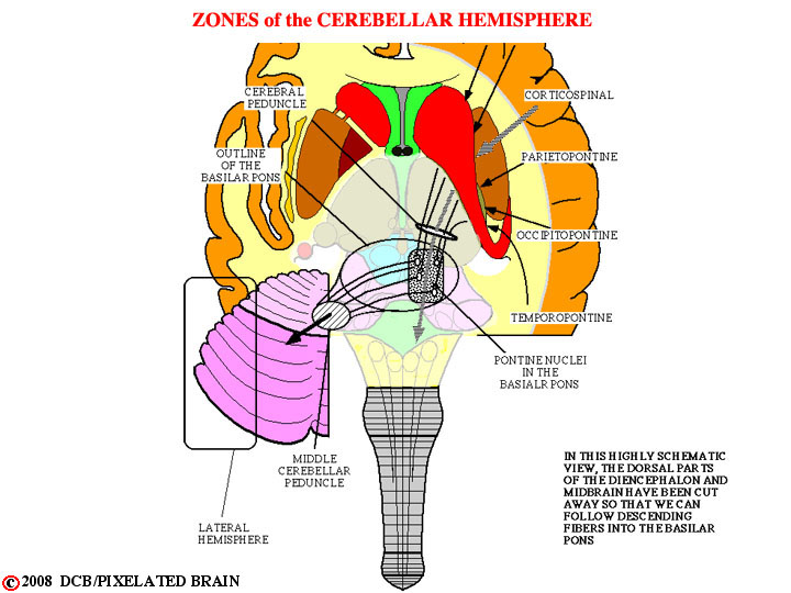
FIGURE 6-7
Perhaps 100 msec. before the onset of a movement neural messages, presumably involving the nature of the desired movement, are sent (at least, potentially) from all four lobes of the cerebral hemisphere to the cerebellum. Axons of the neurons involved pass successively through the corona radiata, the internal capsule and the cerebral peduncle to reach the basilar pons. It is a massive projection and - as with most pathways of this sort - it is orderly in nature. Fibers from the frontal lobe (the fronto-pontine pathway) pass through the anterior limb of the internal capsule and then through the medial one fifth of the cerebral peduncle to reach the basilar pons. Fibers from the remaining three lobes pass through the posterior limb of the internal capsule and the lateral one fifth of the cerebral peduncle to reach the same destination. Within the cerebral peduncle these two groups of fibers are separated by the corticospinal tract, occupying the middle three fifths of the peduncle.
Within the basilar pons the descending fibers from all four lobes terminate by synapsing upon the cell bodies of the nuclei scattered diffusely throughout this region - the so-called pontine nuclei. These neurons relay information to the cerebellum by giving rise to axons which cross the midline, run through the middle cerebellar peduncle and make synaptic contact with granule cells in the cortex of the lateral part of the cerebellar hemisphere. Because of their appearance these axons are called mossy fibers and their specialized region of contact with granule cells is termed a glomerulus,
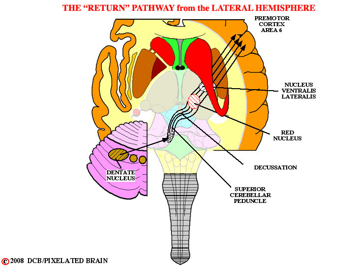
FIGURE 6-8
After neural processing of this input at the level of the cerebellar cortex, purkinje cells relay the output to the dentate nucleus, the largest of the "deep" nuclei, buried within the white matter of the cerebellar hemisphere, as shown here. This output is entirely inhibitory, and thus acts by modulating the tonic activity of neurons within the dentate nucleus.
Neurons within the dentate nucleus have axons which begin the return route to the the cerebral cortex. These fibers exit from the cerebellum in the superior cerebellar peduncle, wrap around the central gray region of the brain stem tegmentum, and cross the midline at the midbrain level. The fibers pass through and around the red nucleus without synapsing and ascend to terminate in the nucleus ventralis lateralis of the thalamus. The thalamic neurons, in turn, relay the message back to the cerebral cortex by way of fibers that pass through the internal capsule.
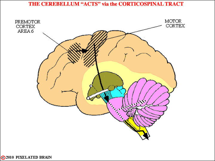
FIGURE 6-9
The next step in this sequence is that the motor cortex, guided in part by information that has been refined by processing in the lateral part of the cerebellar hemisphere, sends motor commands to the spinal cord (and motor nuclei in the brainstem). As shown here, the descending axons of this pathway give off collaterals as they pass through the basilar pons and these fibers synapse on neurons which in turn project through the middle cerebellar peduncle to terminate in the vermis and intermediate region of the cerebellar hemisphere (on the side contralateral to the one where the motor commands originated). Thus, these regions of the cerebellar cortex receive a "copy" of the motor commands being sent to the spinal cord and motor nuclei of the brainstem.
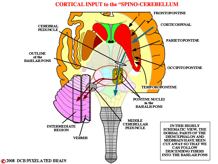
FIGURE 6-10
_ _ _The next step in this sequence is that the motor cortex, guided in part by information that has been refined by processing in the lateral part of the cerebellar hemisphere, sends motor commands to the spinal cord (and motor nuclei in the brainstem). As shown here, the descending axons of this pathway give off collaterals as they pass through the basilar pons and these fibers synapse on neurons which in turn project through the middle cerebellar peduncle to terminate in the vermis and intermediate region of the cerebellar hemisphere (on the side contralateral to the one where the motor commands originated). Thus, these regions of the cerebellar cortex receive a "copy" of the motor commands being sent to the spinal cord and motor nuclei of the brainstem.
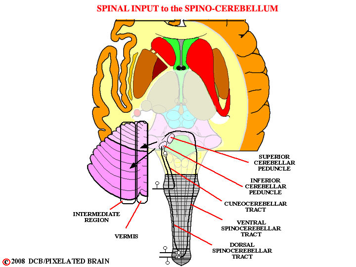
FIGURE 6-11
Once the motor commands have reached the spinal cord and movement has ensued, large diameter afferent fibers from muscle spindles and joint receptors measure the displacement that has actually taken place. This proprioceptive information is relayed to the spinocerebellum by the pathways shown here. This part of the cerebellum makes a comparison between the intended and actual movement. If there is a discrepancy between the two, corrective instructions are sent to the spinal cord.
The output of the intermediate region projects to the red nucleus by way of the superior cerebellar peduncle. Most of the fibers terminate there. Cells within the red nucleus then relay this information down to the cord in the rubrospinal tract. As Kingsley notes, some of the fibers in the superior cerebellar peduncle pass through the red nucleus and continue on, to terminate in the the thalamus. So the information concerned with the adjustment of ongoing motor activity is passed "up" to the cortex as well as "down" to the spinal cord.
The output of the vermis projects to the vestibular nuclei, and on to the cord in the vestibulospinal tracts.
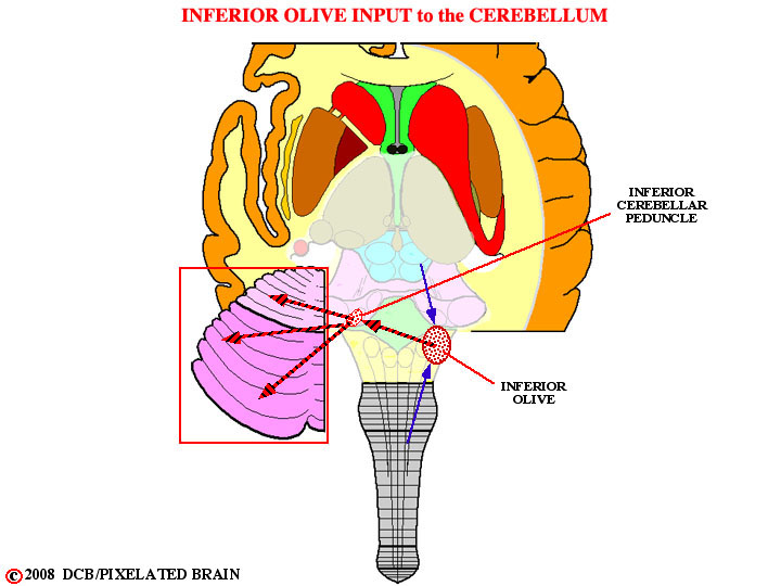
FIGURE 6-12
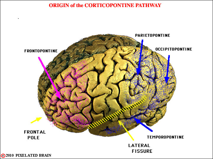
FIGURE 6-13
This view shows the broad origin of the cortical input to cerebellum. The descending pathway from the frontal lobe is colored violet in subsequent views and that from the other lobes is colored dark blue.
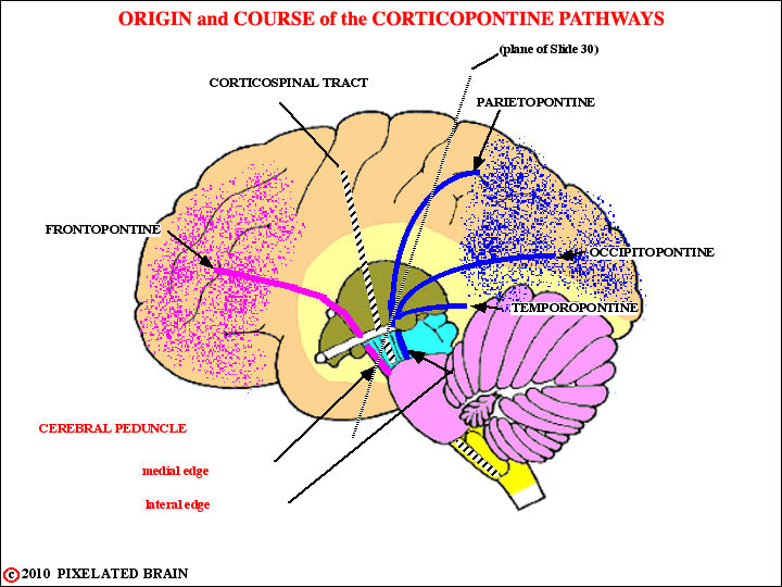
FIGURE 6-14
This view demonstrates how descending corticopontine fibers find their way into the medial 1/5 and the lateral 1/5 of the cerebral peduncle. Note the plane of Slide 30, which will be our next view.
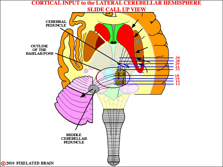
FIGURE 6-15
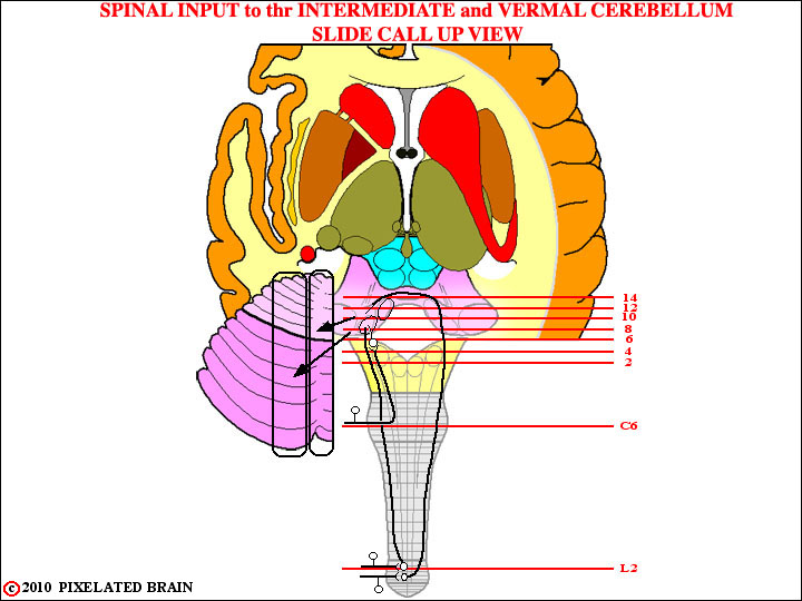
FIGURE 6-16
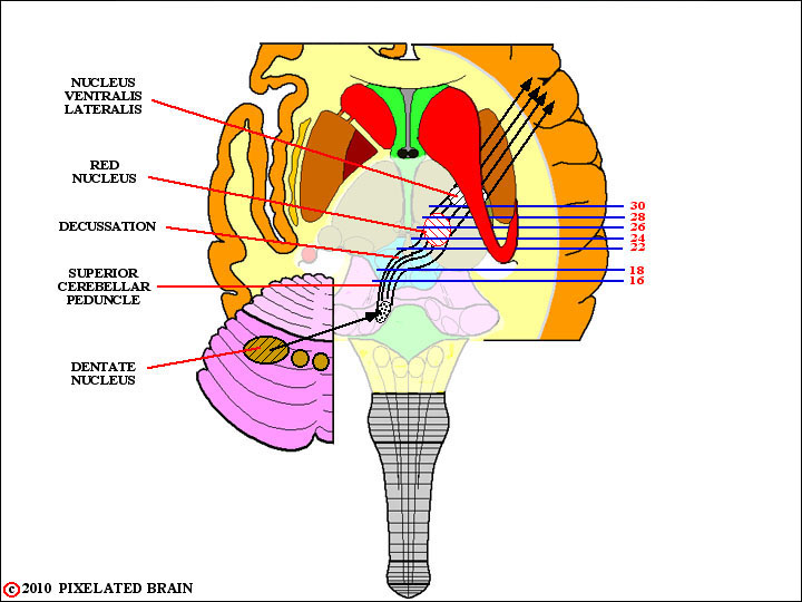
FIGURE 6-17
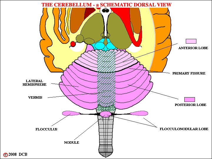
FIGURE 6-18
FIGURE
z
FIGURE
z