MODULE 8 - SECTION 1 - THE CONCEPT OF FUNCTIONAL COMPONENTS
Most of you will be familiar with the concept of functional components from Gross Anatomy.
While we generally avoid dealing with the peripheral nervous system, leaving this for the gross anatomists, it is difficult to do so in the case of the cranial nerves. For example, you can't really understand how to test the accessory nerve unless you are familiar with the actions of the muscles it innervates.
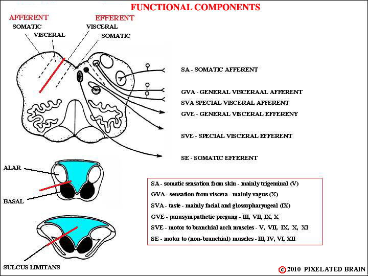
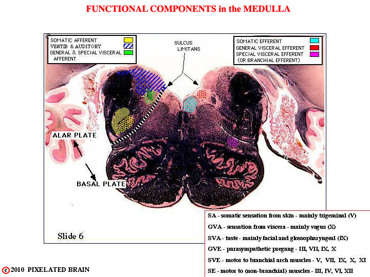
You will remember (we hope) that when the neural plate rolled up to form a tube the sulcas limitans served to form a marker, seperating the dorsal "alar plate" from the ventral "basal plate". There are, however, regions where the lips of the neural plate don't meet; in these regions the roof of the central cavity is sealed by choroid plexus tissue, and the alar plate tends to lie lateral to the basal plate, rather than dorsal to it - as shown above. The picture is further complicated by the appearance of four new functional groupings, serving functions unique to the head. These are thought of as "visceral" because the are associated with the rostral end of the gut or alimentary canal. As the diagrams show, the sensory nuclei lie dorsolateral to the sulcus limitans and the motor nuclei lie ventromedial to it.
The sulcus limitans is only obvious in the caudal medulla, but a suggestion of it is also present in the diencephalon where a slight "valley" separates the dorsal thalamus (which might be thought of as a sensory structure) from the ventrally placed hypothalamus (which might be thought of as a motor structure).
This view shows our color code for the cranial nerves.
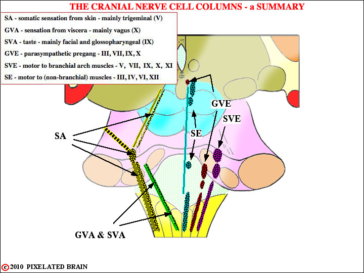
Most books describe the the vestibular and auditory nuclei as part of a "special somatic afferent (SSA)" cell column, grouping them with olfactory and optic structures. - see, for example Blumenfeld's Table 12.4 on Page 470. This doesn't seem like a logical grouping to us, so we drop the SSA category and refer to trigeminal sensory structures as simply "somatic afferent." Unfortunately, we have not been entirely consistent in this and the terms SSA and GVA do creep into some figures.
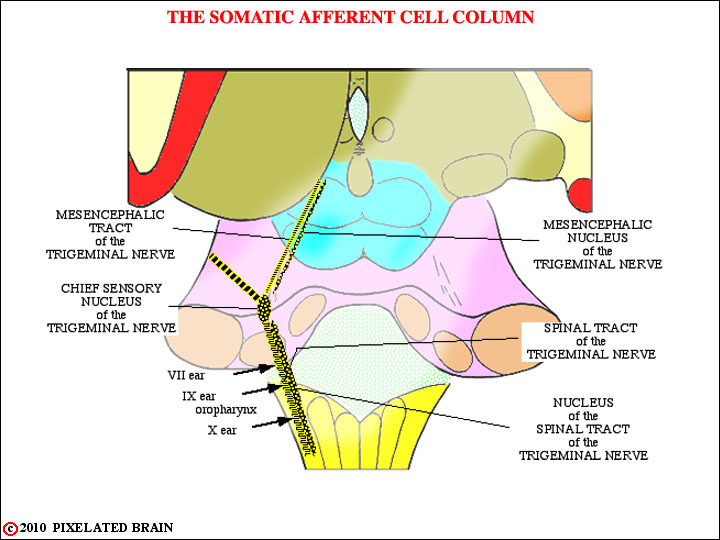
(General) somatic sensory function for the face is served primarily by the trigeminal nerve and we considered these structures in detail in Module 4. The view here is just a schematic version of Figure 4-4.
Something we didn't tell you earlier is that during development all the branchial arches share a common turf in the region aroind the ear. Thus cranial nerves 7, 9 and 10 each have a few somatic sensory fibers. For more detail, see descriptions of the individual nerves, later in the module.
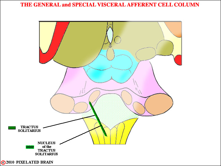
Axons of cranial nerves VII, IX and X conveying the sense of taste (SVA) enter the brainstem within their respective nerves and travel upward in the Tractus Solitarius to terminate in the most rostral part of the Nucleus of the Tractus Solitarius, sometimes known as the gustatory nucleus. Second order neurons within the nucleus send fibers rostrally to terminate in the VPM nucleus of the thalamus. The pathway is probably bilateral, and runs near the ventral secondary trigeminal tract (Blumenfeld places it in the central tegmental tract). From the thalamus, the pathway ascends to a cortical region terminating laterally, near the tongue area of the postcentral gyrus and adjacent to the insula.
Axons of cranial nerves IX and X conveying general sensation (GVA) from the viscera enter the brainstem and travel downward in the Tractus Solitarius to terminate in the caudal part of the nucleus. We are not consciously aware of these sensations. Neurons in this part of the nucleus send axons to terminate in a broad array of brainstem nuclei (the Dorsal Motor Nucleus of the Vagus, the Nucleu Ambiguus, the Reticular Formation) , thus participating in a variety of visceral and autonomic reflexes. Fibers also pass rostrally to provide an input to the Hypothalamus and Amygdala.
Most books do not include the parabrachial nucleus in the taste pathway. It lies adjacent to the superior cerebellar peduncle (Brachium conjuntivum - hence, the name) in the rostral pons.
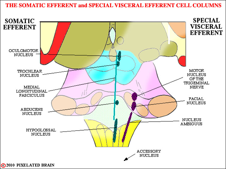
The oculomotor, trochlear and abducens nerves innervate the muscles that move the eyes. The medial longitudinal fasciculus serves to connect these nuclei and coordinate their activity. The hypoglossal nerve, of course, has nothing to do with eye movement - rather, it innervates the muscles of the tongue. These nuclei are all a part of the Somatic Efferent (SE) cell column and we take them up in Module 9
The four nucle of the Special Visceral Efferent (SVE) cell column are shown here in purple. The (spinal) Accessory nucleus lies in the upper 5 or 6 segments of the cord, indicated here by the arrow. While most of the cell columns, like the SE one above are at best, interrupted ones, the SVE column is really an almost continuous one. The motor neurons in these nuclei are typical "lower motor neurons", just like those in the spinal cord. What makes them "special" is that they innervate muscles of branchial arch origin. The muscles are striated, so many people are uneasy about calling them "special VISCERAL motor. To get around this they are often called Branchiomotor.
Just like spinal motor neurons, the SVE ones are driven by upper motor neurons in the lateral part of the precentral gyrus. This pathway departs from the corticospinal one at the midbrain level (Figure 5-5), so we can't call it part of the pyramidal tract (it doesn't go through the pyramids of the medulla). It is called the coriticobulbar pathway and you traced it in Module 5 (remember all those yellow dots). If you want a quick review, click on Slide 28, and then use the "caudal buttons" to follow it downward through the tegmentum. We discuss it later in the module.
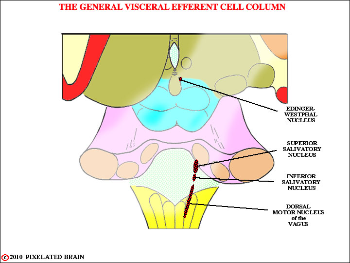
As you know, the autonomic nervous system is made up of two parts - the sympathetic (thoracolumbar) system and the parasympathetic (craniosacral). Both systems have 2 neuron pathways , involving preganglionic and postganglionic neurons. Here , we are looking at the cranial part of the parasympathetic nervous system, and the nuclei you see contain preganglionic neurons. The neurons in the Superior Salivatory Nucleus will send axons out in the Facial Nerve: the ones in the Inferior Salivatory Nucleus contribute axons to the Glossopharyngeal Nerve, and the ones in the Dorsal Motor Nucleus of the Vagus contribute axons to the Vagus Nerve.
We shouldn't forget the Nucleus of Edinger-Westphal. It contributes parasympathetic axons to the oculomotor nerve... yes, it's not a branchiomeric nerve, but it does have parasympathetic fibers in it. Go figure!
For more information about these GVE (parasympathetic) fibers, see the views of the individual cranial nerves.
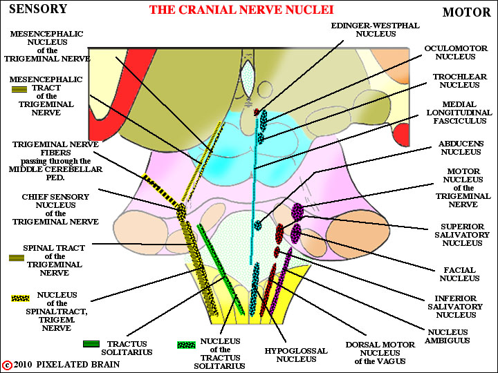
Well, we didn't want to show you this view at the start, because we figured you'd never get past the first slide. Most books have views of this sort. The thing that makes this view special is that it was constructed to scale from a set of slides cut through a real brainstem ....the slides we call the Cu Series. You can check it out by calling up FIGURE 8-2 and looking at specific slides in the series.