MODULE 8 - SECTION 2 - THE POSITION of the FUNCTIONAL CELL COLUMNS
in
SECTIONS through the BRAINSTEM
Before we begin to look at individual slides, it may help to get an overview of the actual nuclei that make up the cell columns.
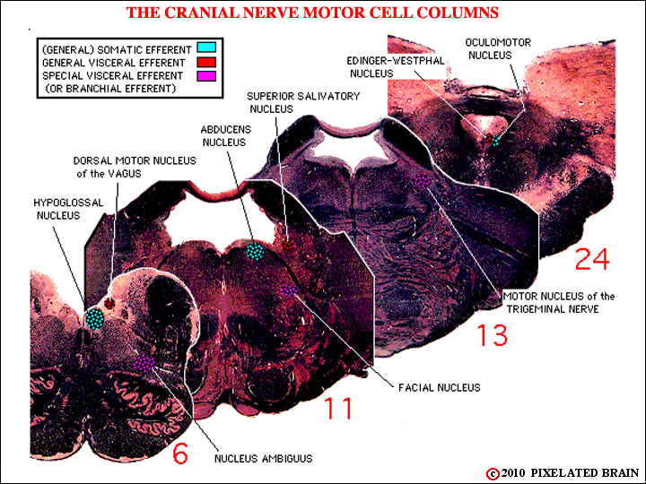
The motor nuclei are shown here.
The idea here was to try to piece together our slides 6, 11, 13, and 24 to demonstrate that the cell columns really are columns, but it isn't easy to do on a limited number of sections. Here we are looking at the motor columns and you may be able to see that:
1) The Nucleus Ambiguus, Facial Nucleus and Trigeminal Motor Nucleus (SVE) do line up, one in front of the other (but difficult to pick out because of the colors used)
2) The Dorsal Motor Nucleus of the Vagus, the Superior Salivatory Nucleus and the Edinger-Westphal Nucleus (GVE) sort of line up, but it is a very discontinuous cell column. (The Inferior Salivatory Nucleus isn't present on the slides selected).
3) The Hypoglossal Nucleus, the Abducens Nucleus and the Oculomotor Nucleus (SE) line up, but again the column is a discontinuous one. (The Trochlear Nucleus isn't present on the slides selected).
Section 4 has the full slide series.
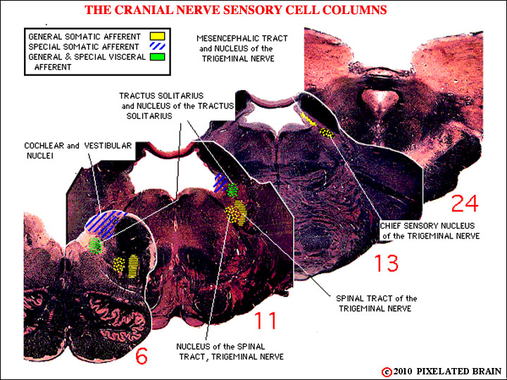
The sensory nuclei are shown here.
This view is of the sensory columns. We have slipped up, as we said we might, and called the Vestibular and Auditory Nuclei "Special Somatic Afferent". Try to ignore this.
The idea here is just to show that the Nucleus of the Tractus Solitarius is a column (admittedly, a short one) that is present in slides 6 and 11 of our slide series (section 4), and that the trigeminal nuclei form a column that extends from the spinal cord (not shown) as far rostrally as slide 13 (and beyond).