MODULE 10 - SECTION 2 - THE ANTERIOR ARTERIAL SUPPLY to the BRAIN
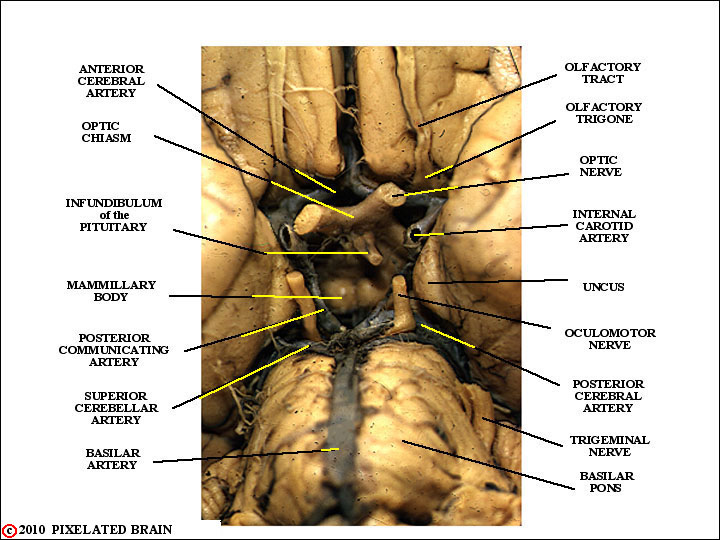
A BASAL VIEW of the HYPOTHALAMIC AREA of a
GROSS BRAIN
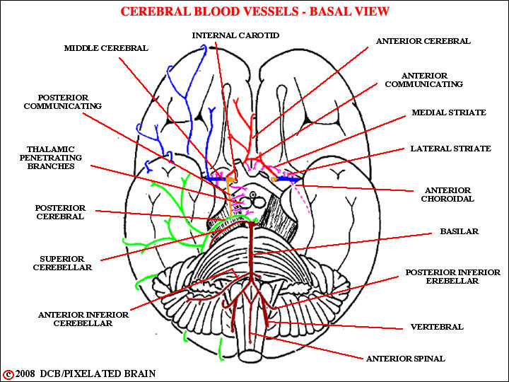
The anterior supply refers to that part derived from the internal carotid arteries. There is a tendency to forget that the internal carotid artery supplies only part (granted, the major part) of the hemisphere. The balance is, of course, supplied by the posterior cerebral artery which is fed by the basilar artery and hence a part of the posterior circulation. This distinction is quite clear to the the neuroradiologist. If they want to visualize the anterior circulation, they inject dye into the internal carotid artery (see Blumenfeld's Figure 4.16); if they want to visualize the posterior circulation they inject dye into the vertebral (see Blumenfeld's Figure 4.17). The internal carotid enters the cranium through the carotid foramen, then turns to run anteriorly and pass through the cavernous sinus where it gives off its first small branch, the ophthalmic artery. It then penetrates the dura to enter the cranial cavity, promptly taking part in the formation of the Circle of Willis- as shown here and in the next figure.
a BASAL VIEW
Another view of the internal carotid.
a MIDSAGITTAL VIEW
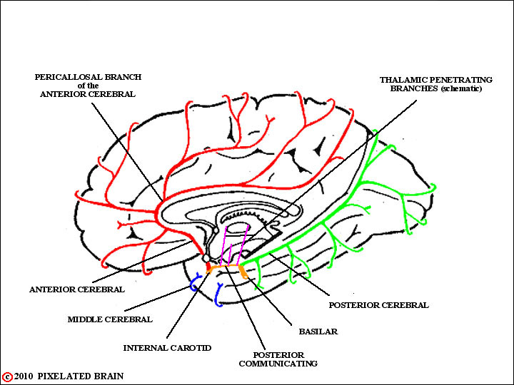
The anterior cerebral artery passes medially, between the base of the brain and the optic nerve, and then rostrally around the genu of the corpus callosum as shown here.
a LATERAL VIEW
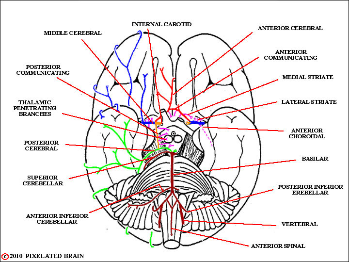
Near their origin the vessels from the two sides are joined by the anterior communicating branch. In the case of the anterior cerebral artery, and all the others we will describe, the important thing for you to remember is the territory supplied by the vessel. Many of the branches of these large vessels have names, but the names differ from one authority to another. So, your best bet is to identify branches in terms of the gyri or regions which they supply. Blumenfeld shows this clearly in his Figures 10.5 and 10.9 The one named branch of the anterior cerebral that's worth remembering is the pericallosal artery. It is actually a continuation of the main trunk of the artery and it is of note because it "hugs" the corpus callosum and serves as a marker for that structure. To confirm this point, find the anterior cerebral artery in Blumenfeld's Figure 4.16C. Equally important, the anterior cerebral vessels are markers of the midline in AP arteriograms. In this case, Blumenfeld's Fig. 4.16A nicely makes the point
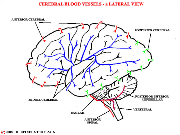
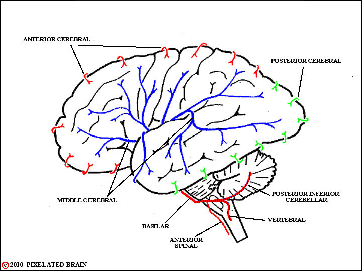
The middle cerebral artery passes laterally between the orbital surface of the frontal lobe and the rostral pole of the temporal lobe, and divides into superior and inferior divisions, as shown in Blumenfeld's Figure 10.6. The inferior division forms several branches which run over the superior temporal gyrus to exit from the lateral fissure and supply the temporal lobe. The superior division breaks up into several branches which ascend over the surface of the insula. These vessels then turn downward, again, to find their way out onto the surface of the brain, where they supply most of the convexity of the frontal, parietal, and occipital lobes. The fact that the branches cover the surface of the insula in such a consistent way means that they are useful markers, defining the position of this buried cortical region, in arteriograms.
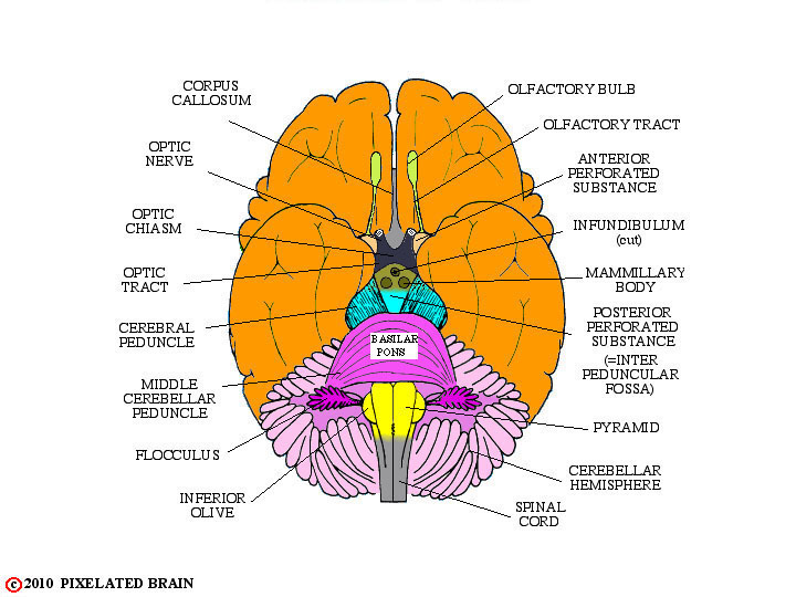
Near their origins, the anterior and middle cerebral arteries give off a series of small blood vessels that rather directly penetrate the brain. Many of them do so in the region between the medial and lateral olfactory stria, causing this area to be termed the anterior perforated substance.
is the SAME at all levels of the
CENTRAL NERVOUS SYSTEM
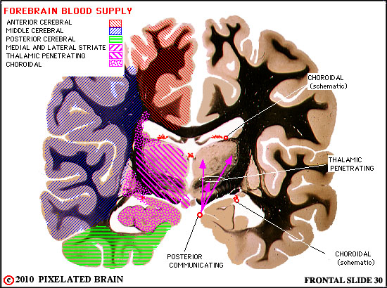
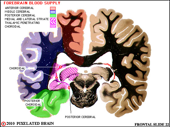
The striates are particularly important because they supply much of the internal capsule . You should also be aware of the anterior choroidal artery (Figure 10-3 at left again) and Blumenfeld's Figure 10.8A); this vessel seems to penetrate the brain, but in fact it follows the optic tract into the sulcus separating the base of the brain from the medial edge of the temporal lobe and ultimately supplies the choroid plexus in the inferior horn of the lateral ventricle. This artery follows the course of the choroid plexus and receives reinforcing branches from the posterior cerebral artery (the posterior choroidal arteries). It is of importance because it also supplies tissue adjacent to the lateral ventricle (Blumenfeld's Figure 10.9A,B and Haines Figure 15-18).
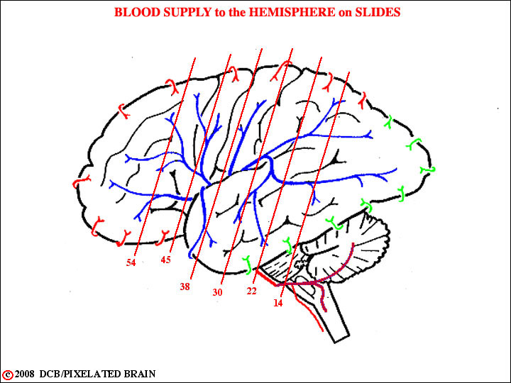
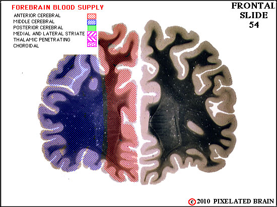
 FOREBRAIN BLOOD SUPPLY
on SLIDE 54

FOREBRAIN BLOOD SUPPLY
on SLIDE 45
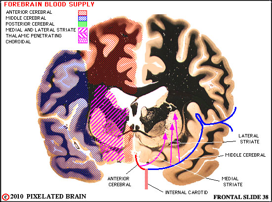
FOREBRAIN BLOOD SUPPLY
on SLIDE 38
FOREBRAIN BLOOD SUPPLY
on SLIDE 30
FOREBRAIN BLOOD SUPPLY
on SLIDE 22
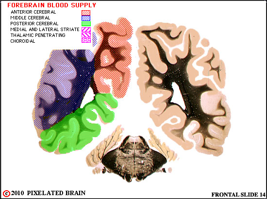
FOREBRAIN BLOOD SUPPLY
on SLIDE 14