MODULE 11 - SECTION 4 - THE OPTIC CHIASM
This small region is of enormous clinical importance - in part because of the reorganization of the visual pathway that takes place here, and in part because of the many structures that lie almost in contact with the chiasm. Let's consider the general anatomy of the region first.
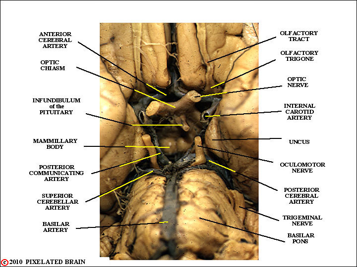
THE HYPOTHALAMIC AREA
on a BASAL VIEW
of the GROSS BRAIN
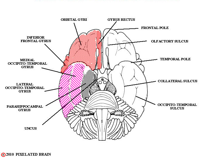
SULCI and GYRI on a BASAL VIEW
of the GROSS BRAIN
This is a drawing of the same basal view.
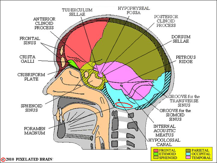
A MID-SAGITTAL VIEW of the SKULL
is a drawing in which the hypophyseal fossa, tuberculum sellae and clinoid processes are labeled.
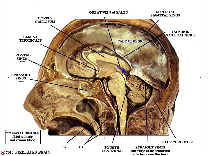
A MIDSAGITTAL VIEW of the BRAIN within the SKULL
Here is the real thing, but in this case the brain has been displaced upward and backward somewhat, so that the relationship of the chiasm and infundibulum to the hypophyseal fossa is distorted.
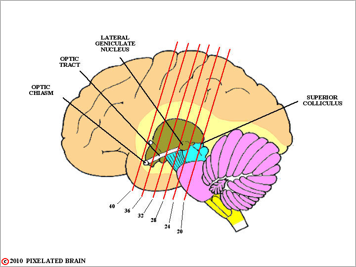
SLIDE LEVELS on a LATERAL VIEW of the BRAINSTEM
From the point of view of the visual system, the important thing that takes place within the chiasm is a "regrouping" of the fibers conveying information to the lateral geniculate nucleus. In essence, the fibers from the nasal half of each retina cross to the contralateral side of the brain in the chiasm, while the fibers from the temporal half of the retina do not, remaining on the ipsilateral side of the brain.
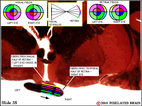
THE VISUAL PATHWAY on SLIDE 38

This slide depicts the fibers from the nasal retina of the left eye crossing in the chiasm. Note the layering of the pathway. This means that pressure on the chiasm from below (a pituitary tumor, perhaps) might cause a somewhat different visual field loss from that caused by pressure from above (a hypothalamic mass, for example).
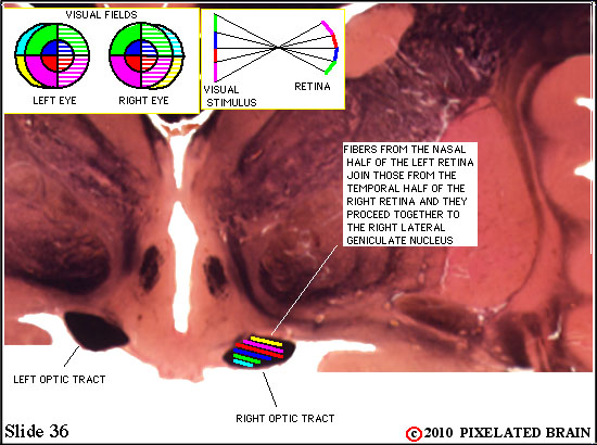
THE VISUAL PATHWAY on SLIDE 36
This slide shows the regrouping that has taken place "behind" the chiasm, within the optic tract.