MODULE 11 - SECTION 5 - THE OPTIC TRACT
The
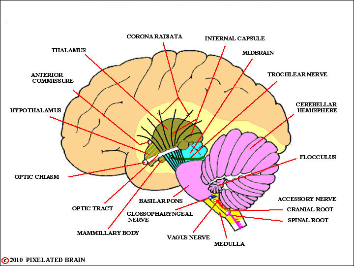
LANDMARKS on a LATERAL VIEW
of the BRAINSTEM
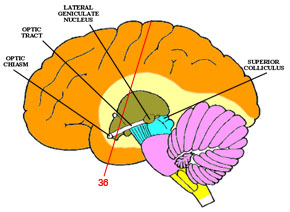
Given all the excitement at the level of the chiasm, this next stretch of the pathway seems rather dull, but also a bit misleading. It seems as though the optic tract is running through the brain, but in fact it runs over the surface of the brain, and has been covered over by the expanding hemisphere. If you cut away the temporal lobe, as was done here , it is clear that the optic tract is actually running over the surface of the cerebral peduncle to reach the lateral geniculate nucleus.
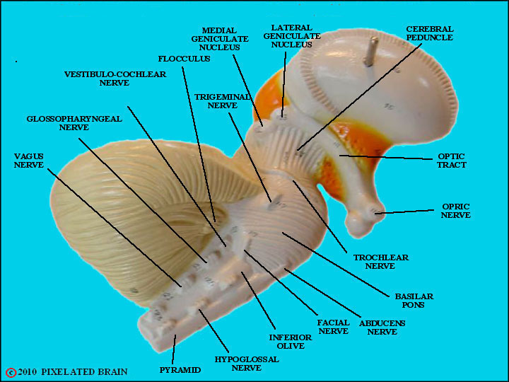
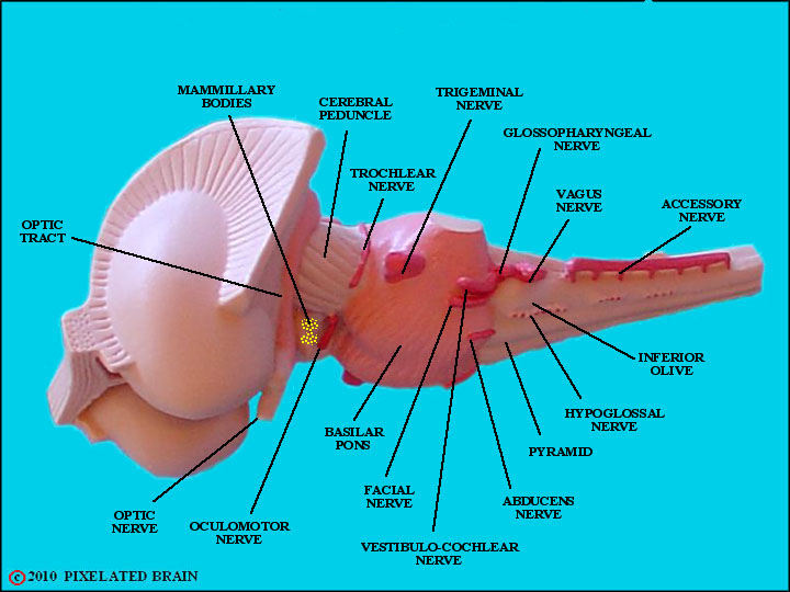
A LATERAL VIEW of the BRAINSTEM on a MODEL
Start at the optic nerve; ignore the fact that the chiasm is vague, so continue to trace the course of the tract. it passes over the surface of the hypothalamus (orange in this view, but pea green in the one above), and then over the cerebral peduncle to reach the lateral geniculate nucleus.
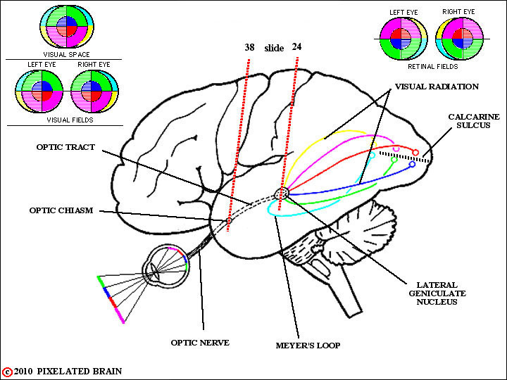
THE VISUAL PATHWAY on a LATERAL VIEW of the BRAIN
Since this slide series starts behind the chiasm, our firs slid will be #36 and we will sintinue caudally to Slide #26.
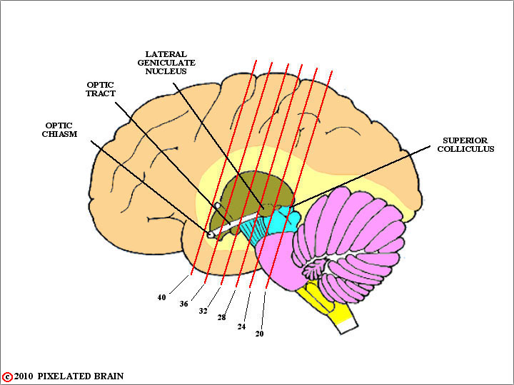
SLIDE LEVELS for the
VISUAL PATHWAY
shown on a LATERAL VIEW of the BRAINSTEM
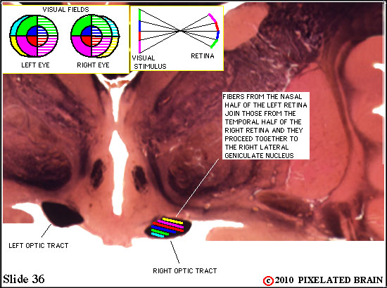
VISUAL PATHWAY on SLIDE 36
X
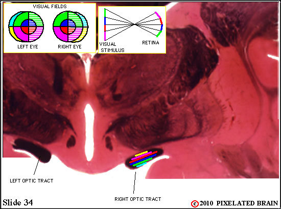
VISUAL PATHWAY on SLIDE 34

X
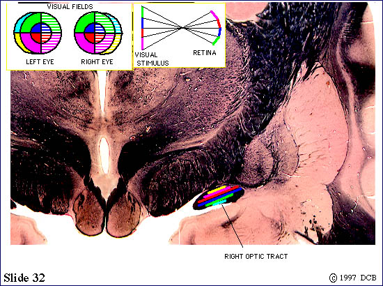
VISUAL PATHWAY on SLIDE 32

X
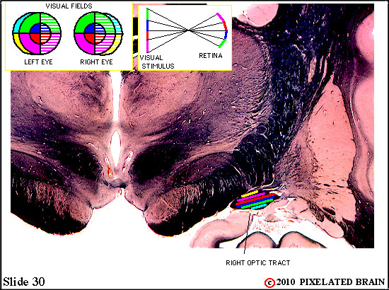
VISUAL PATHWAY on SLIDE 30

X
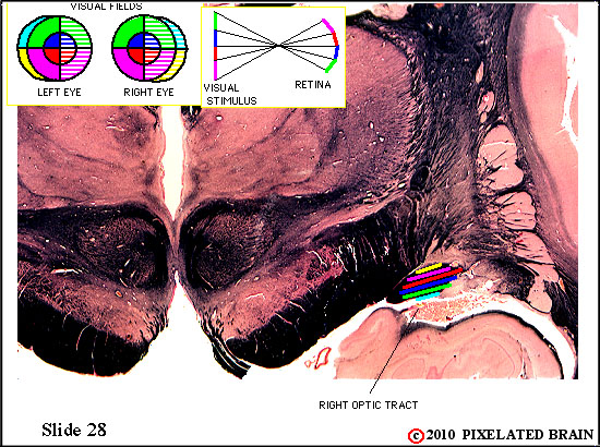
VISUAL PATHWAY on SLIDE 28

X
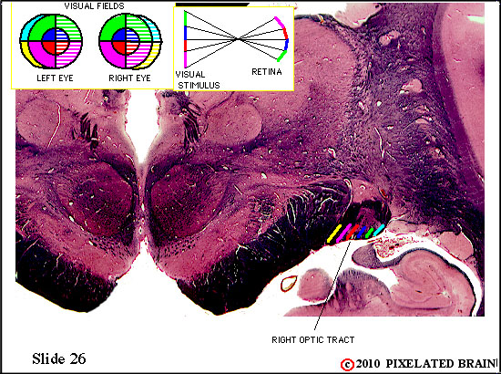
VISUAL PATHWAY on SLIDE 26

Some would say that the optic tract is now beginning to rotate; for the right tract, shown here, it is a 90 degree counter-clockwise rotation, such that the yellow fibers move from the "top" of the tract to the medial edge as they prepare to enter the most medial pary of the lateral geniculate nucleus.
THE BRAINSTM
an OBLIQUE LATERAL VIEW
Now, follow the tract on this model.
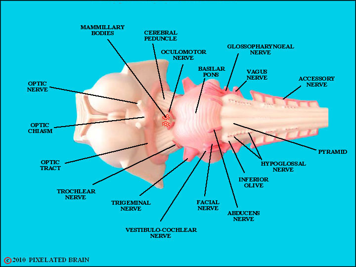
THE BRAINSTEM - a BASAL VIEW
This ventral view of the same model shows the optic chiasm and tract.
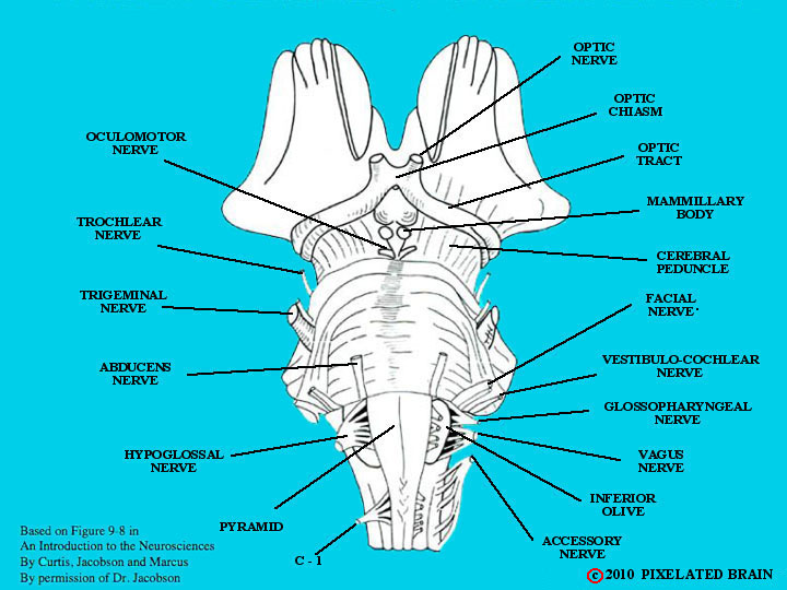
THE BRAINSTEM - a BASAL VIEW
And here, another basal view.
13