MODULE 14 - SECTION 1 - THE DIENCEPHALON - HOW IT DEVELOPED
and
WHAT IT LOOKS LIKE
This section is a review of material that has been covered in earlier modules, so you can skip it if you think you are familiar with the subject
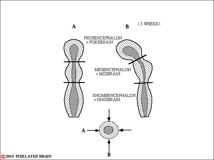
THE THREE PRIMARY VESICLES
Recall that the brain starts out as a simple tube-like structure.
EARLY DEVELOPMENT of the BRAIN
Within the cranial cavity, the more caudal part of the tube develops into the medulla, pons and midbrain. Collectively these regions are known as the brain stem. The most rostral division of the neural tube, the forebrain, develops into the diencephalon and telencephalon (or cerebral hemisphere).
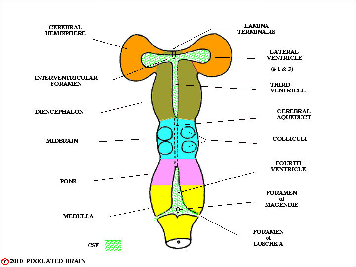
The color code used in this view will be used in the figures that follow, which trace the development of the brain.
Cerebral Hemisphere orange
Diencephalon olive green
Midbrain blue
Pons (and Cerebellum) purple
Medulla yellow
DIVISIONS of the BRAINSTEM
These regions are complex and may be subdivided into several components.
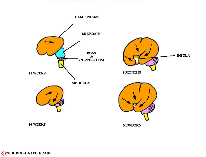
THE EXPANDING HEMISPHERE
In this module we will start by considering the diencephalon as a whole, but then focus on the dorsal thalamus which - from this point on - we will refer to simply as the thalamus. Views such as the previous one above would suggest that we ought to be able to see the diencephalon "from above". But, because of the way in which the hemispheres expand to cover the more caudal part of the brain the diencephalon is hidden from view both dorsally and laterally.
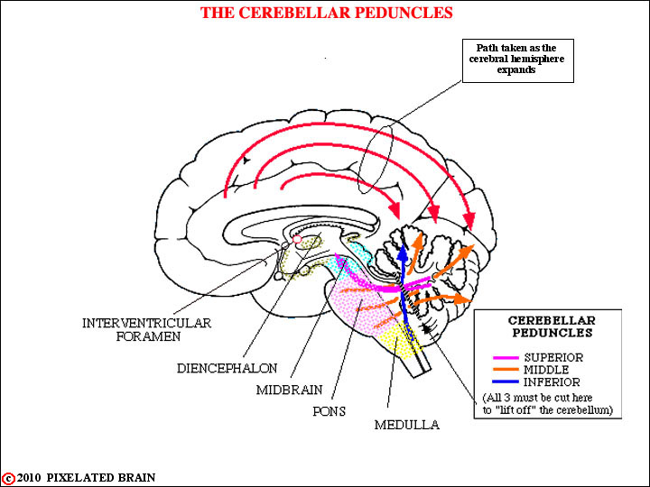
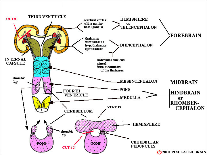
THE CEREBELLAR PEDUNCLES
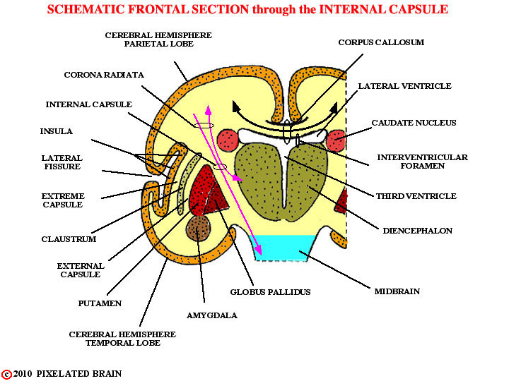
SCHEMATIC FRONTAL SECTION
through the
INTERNAL CAPSULE
The diencephalon is surrounded, both dorsally and laterally, by the expanding cerebral hemisphere.
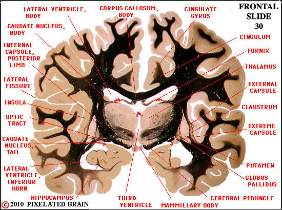
ATLAS VIEW - FRONTAL SLIDE 30
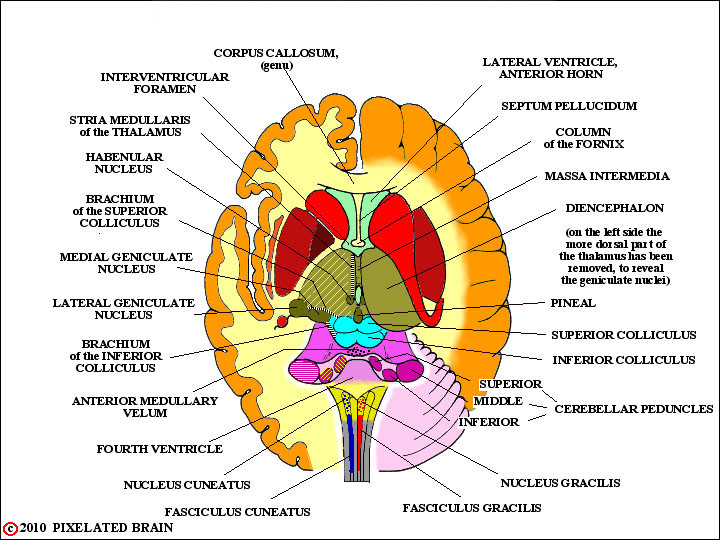
This atlas view, and the two that follow, simply relates the diencephalon (thalamus) to surrounding structures.
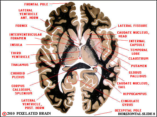
ATLAS VIEW - HORIZONTAL SLIDE 30
This atlas view simply relate the diencephalon (thalamus) to surrounding structures.
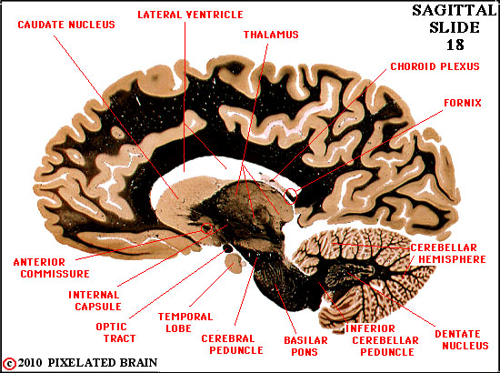
ATLAS VIEW - SAGITTAL SLIDE 18
This atlas view simply relates the diencephalon (thalamus) to surrounding structures.
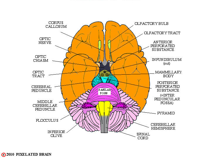
A BASAL VIEW of the BRAIN
Only a very limited part of this diencephalon - the hypothalamus - can actually be viewed without removing brain tissue. The surface of the hypothalamus is colored olive-green in the view, but only one landmark (the mammillary body) is labeled.
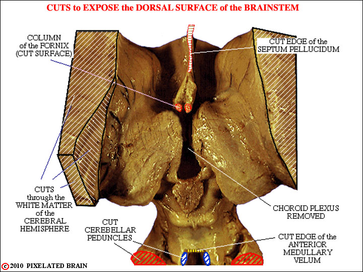
HOW to EXPOSE the THALAMUS - 1
If we had the luxury of working with real brains, we would expose the dorsal surface of the thalamus by cutting away the overlying hemisphere. In conceptual terms, we would make cut #1 as shown on the left on both sides and lift off the hemispheres.
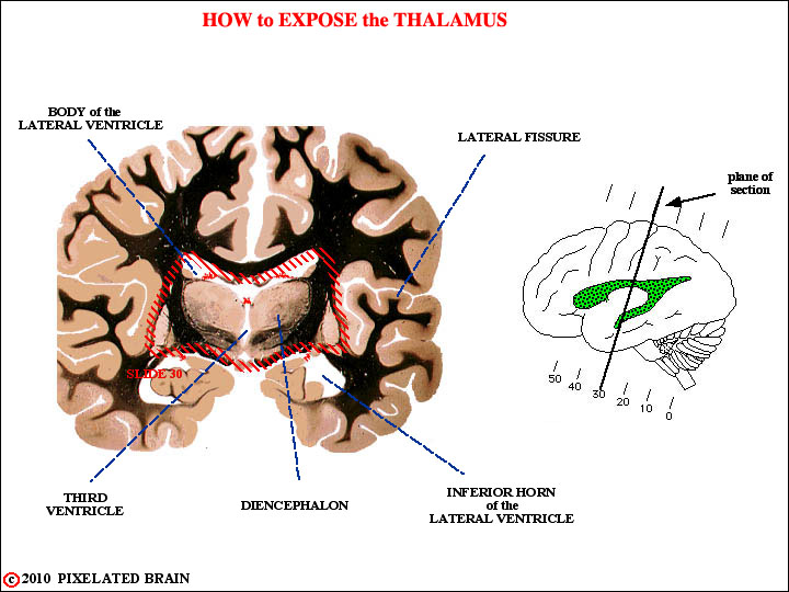
HOW to EXPOSE the THALAMUS - 2
This section shows the brain before such a cut.
12

HOW to EXPOSE the THALAMUS - 3
This section shows the brain after the cut.
THE THALAMUS EXPOSED - 1