PIXBRAIN HOME _ _ MOD 2 HOME _ _ previous _ _ FIGURE 2-1 _ _ next _ _ I WANT TO
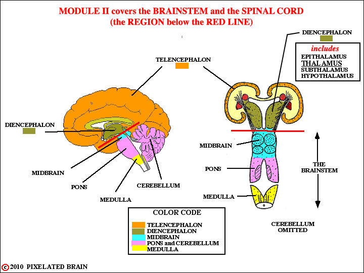
- - As you will recall, we covered the anatomy of the Forebrain in Module 1. In Module 2 we will deal with the Brainstem and Spinal Cord. Because we didn't spend much time on the Diencephalon in Module 1, and because it has a close functional relationship with the Brainstem, we will include it in our discussions. The box in the upper right corner of this view lists the components of the Diencephalon. We have printed "Thalamus" in larger type to remind you that it is by far the largest division of the Diencephalon.
- - The brain is, from an embryological point of view, the rostral end of the neural tube - continuous caudally with the spinal cord in the region of the foramen magnum. Unlike the spinal cord, in which each segment looks more or less like the next one, regional differences in development give each division of the brainstem its own peculiar appearance. The next slide provides some of these details.
- - In the drawings that follow we will do our best to stick to the color scheme given in the lower left corner of this view.