PIXBRAIN HOME _ _ MOD 1 HOME _ _ previous _ _ FIGURE 1-9 _ _ next _ _ I WANT TO
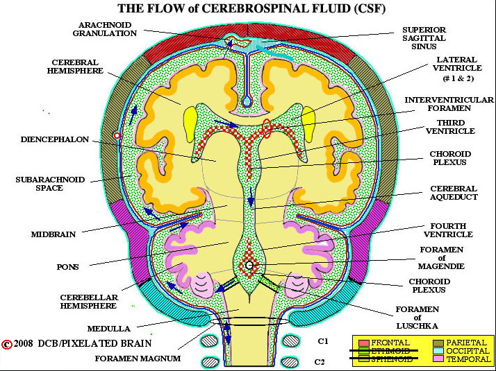
- - This viewtraces the flow of CSF. From the major site of formation, in the two lateral ventricle, fluid passes through the interventricular foramen to enter the third ventricle. A bit more fluid comes from the choroid plexus roofing the third ventricle; CSF exits into the cerebral aqueduct and descends into the fourth ventricle. Again, choroid plexus in the roof of this ventricle contributes a small amount of CSF to the total. CSF exits from the ventricular system through paired lateral openings, the Foramina of Luschka, and a single midline opening in the roof of the ventricle, the Foramen of Magendie. Once outside, the fluid lies within the subarachnoid space - a region defined more precisely in a coming view. For now, just trace the arrows up over the surface of the brain to the arachnoid granulation. CSF enters these specialized structures and from here returns to the venous blood of the superior sagittal sinus.
- - Diagrams of choroid plexus tissue (Blumenfeld Fig. 5-13, Fitzgerald Fig. 5-17, Haines Fig. 6-18) show that the fenestrated capillaries within choroid tissue allow for free passage of water and solutes but that tight junctions between choroid epithelial cells form a blood-CSF barrier. While lipid soluble substance may easily pass through this barrier, most other materials must be conveyed from blood to CSF by active transport through cells of the choroid epithelium. The result is that the chemical composition of the two fluids differ; for example, CSF has higher levels of chloride, magnesium and sodium, compared with plasma. CSF is colorless, low in protein and contains very few, if any, white blood cells.
- - The volume of CSF in the adult is about 120mL and roughly 500mL of the fluid is produced every day, so that about 4 turnovers occur in 24 hour. Production continues even if there is a blockage of flow, somewhere along the way to the arachnoid granulations, but the result depends on the age of the subject and the site of the obstruction. For details, see Blumenfeld, pages 151-2/ Fitzgerald, pg. 51/ Haines, pg. 101)
- - Most CSF returns to the vascular system by entering arachnoid granulations. Some fluid passes between the cells lining a granulation to mix with the venous blood of the superior sagittal sinus. Most of the CSF, however, is transported through the cells in membrane bound vesicles. Granulations are easily seen in gross specimens. If the dura is carefully removed from the surface of the brain, they tend to pop out of the superior sagittal sinus and be exposed to view, as in Figure 1-13. Another approach is to open the superior sagittal sinus in the midline and look at the granulations as they protrude into the sinus (Figure 1-14).