PIXBRAIN HOME _ _ MOD 2 HOME _ _ previous _ _ FIGURE 2-10 _ _ next _ _ I WANT TO
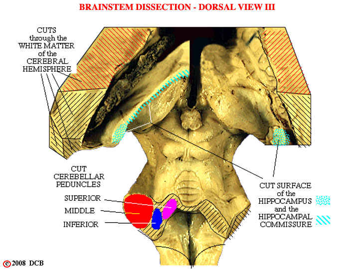
- - This dissection is similar to that of Figure 2-9, but photographed at a slightly different angle. It shows the cut cerebellar peduncles and the midbrain more clearly, but gives a more oblique view of the diencephalon. In this instance part of the fornix and related structures (the hippocampus and hippocampal commissure - don't worry about them, for now) were left in place on the left side, partially obscuring the dorsal surface of the diencephalon.