PIXBRAIN HOME _ _ MOD 2 HOME _ _ previous _ _ FIGURE 2-11 _ _ next _ _ I WANT TO
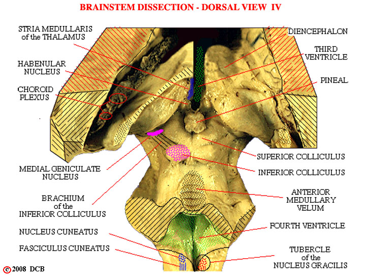
- - This view identifies a variety of structures. Features you should note include:
1) the choroid plexus. You should realize that our dissection has exposed much of the lateral ventricle, as well as the dorsal surface of the brainstem . The choroid plexus remained behind, when the hemisphere was removed, and can be seen passing around into the inferior horn of the ventricle, just as shown in Figure 2-9.
2) the diencephalon. The more caudal part of the diencephalon tends to "overhang" the midbrain and may obscure it from view . In Figure 2-5, this part of the diencephalon has been shown on the right but "removed" on the left to reveal underlying structures.
3) the brachium of the inferior colliculus. One of the many bundles of fibers which happen to run on the surface of the brainstem and therefore create an identifiable surface landmark. Brachium is one of several names used for structures of this sort. You may want to look at Figure 2-12 for more examples of terms we will begin to use.
4) the superior, middle and inferior cerebellar peduncles. Here we see the cut surface of the peduncles. They are, in a sense, embedded in surrounding tissue, and thus their borders are not easily defined in the photograph; they are shown more precisely in Figure 2-5.
5) the fourth ventricle. It is exposed to view because the cerebellum and choroid plexus have been removed.
6) the nucleus cuneatus, fasciculus cuneatus and tubercle of the nucleus gracilis. These structures lie at the most caudal level of the brainstem, where the medulla becomes continuous with the spinal cord. The tubercle is simply the surface bump, created by the underlying nucleus.