MODULE 4 - THE ANTEROLATERAL PATHWAY
The impo
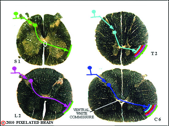 Figure 0
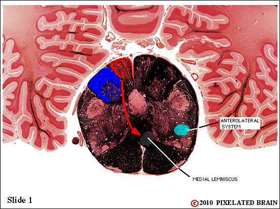 Figure 1
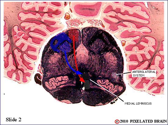 Figure 2
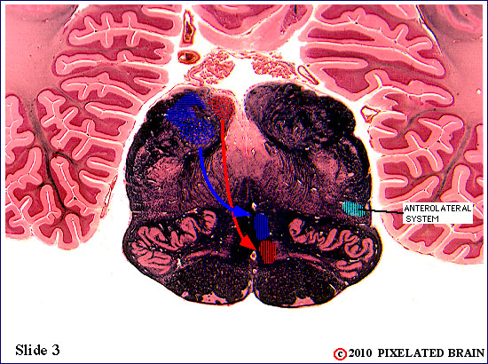 Figure 3
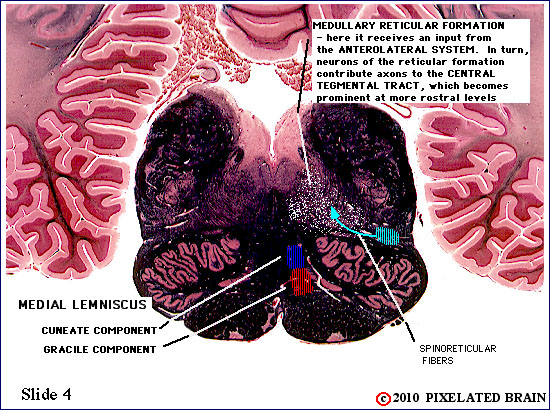 Figure 4
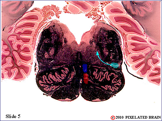 Figure 5
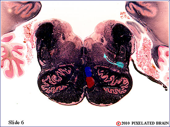 Figure 6
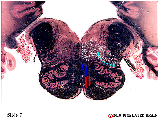 Figure 7
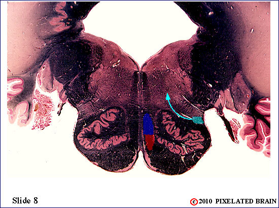 Figure 8
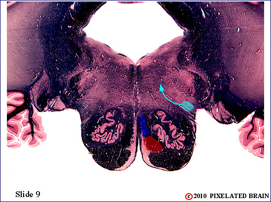 Figure 9
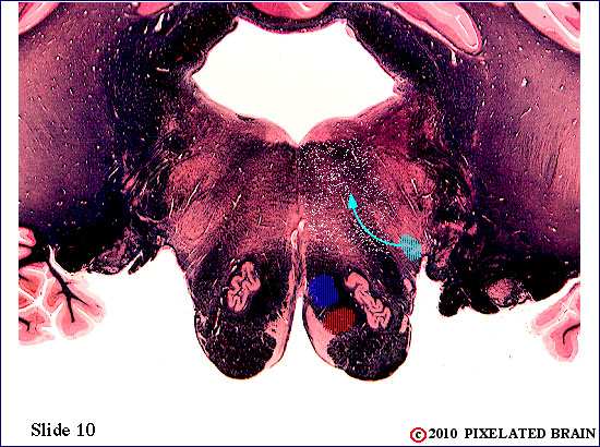 Figure 10
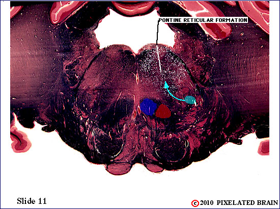 Figure 11
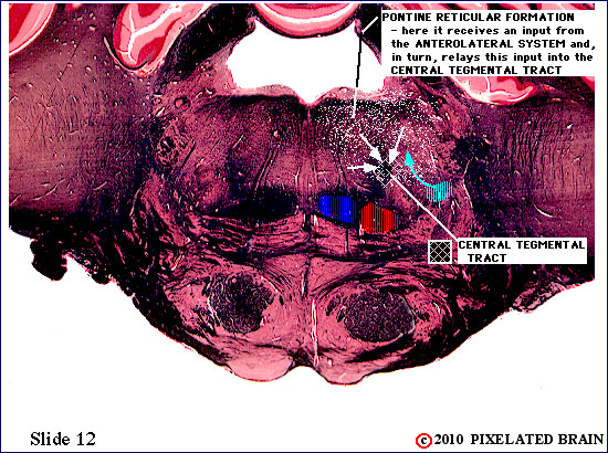 Figure 12
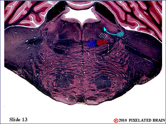 Figure 13
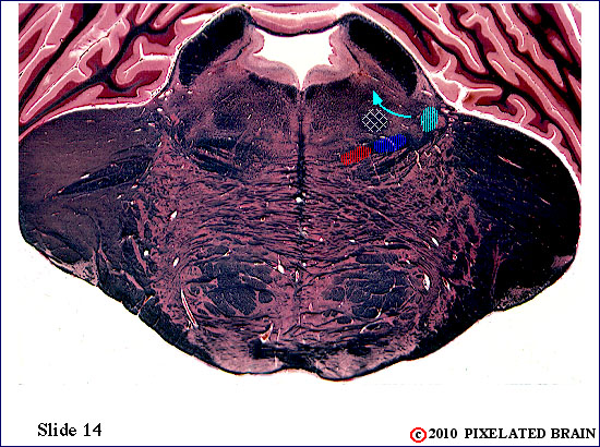 Figure 14
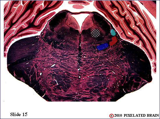 Figure 15
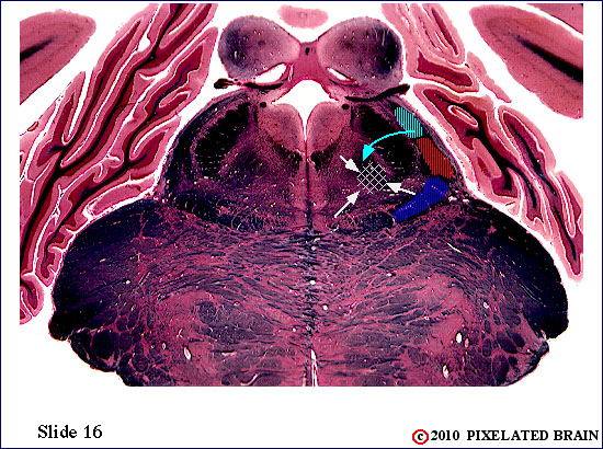 Figure 16
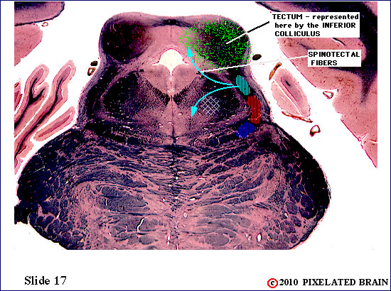 Figure 17
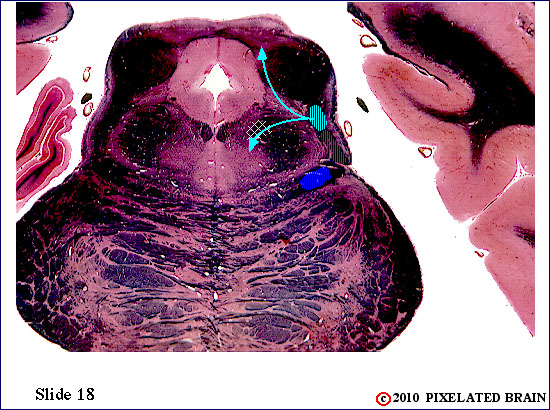 Figure 18
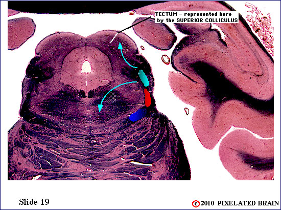 Figure 19
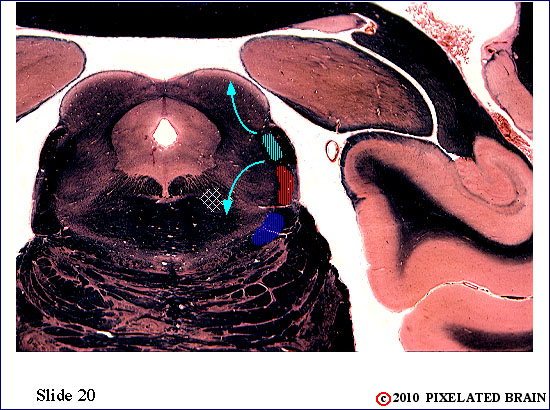 Figure 20
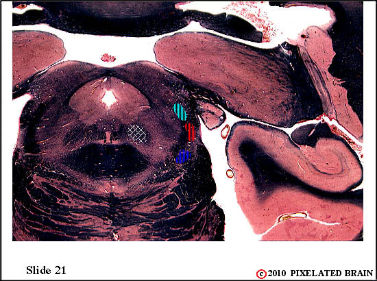 Figure 21
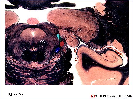 Figure 22
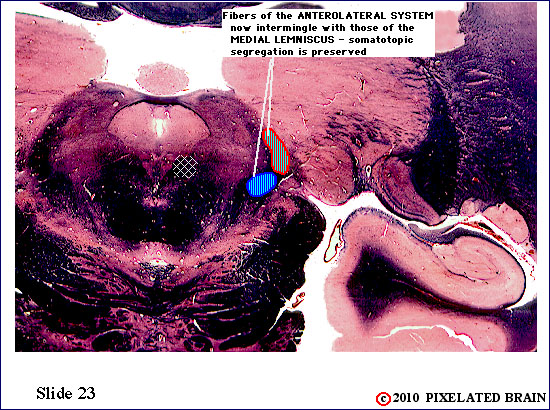 Figure 23
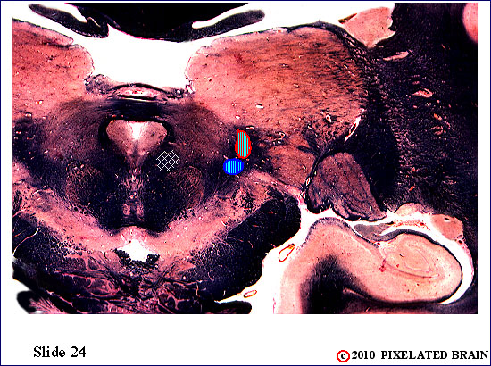 Figure 24
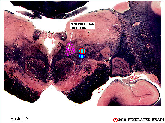 Figure 25
 Figure 26
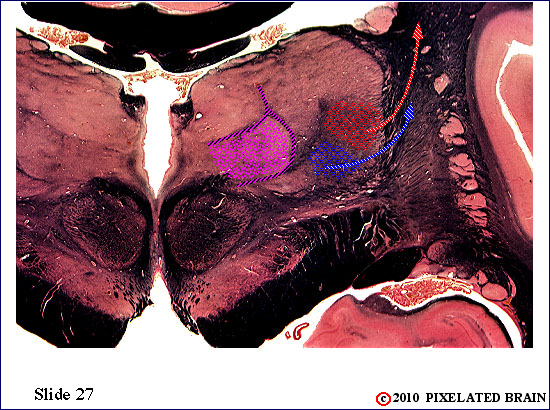 Figure 27
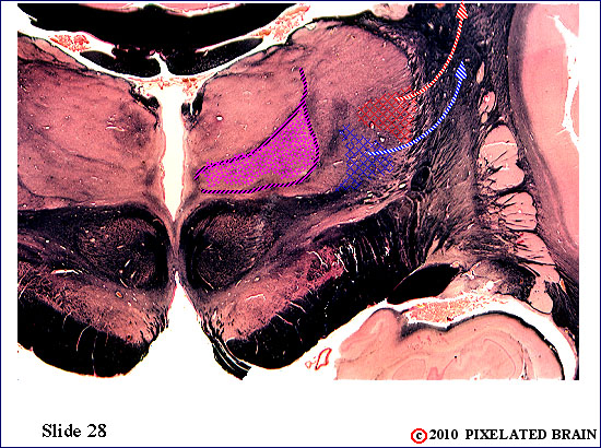 Figure 28
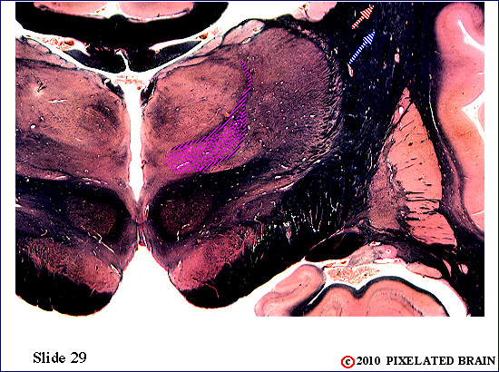 Figure 29
Fibers of this pathway are organized in a somatotopic manner (fibers from adjacent parts of the body's surface run in adjacent parts of the pathway), and the color code used to represent this is:
- Sacral - green
- Lumbar - purple
- Low Thoracic - red
- High Thoracic - light blue
- Cervical - dark blue
The first order neurons of this pathway are large myelinated axons which enter in the medial part of the dorsal root. The region where each dorsal root enters the spinal cord is often called the "dorsal root entry zone" and these fibers enter in the medial part of the zone. The fibers then bifurcate, with one branch passing deeply to terminate in the dorsal horn and the other branch passing medially to ascend in the posterior funiculus. Because of the way in which the ascending fibers pile up in the posterior funiculus, we say that it is somatotopically organized, with the lower part of the body represented most medially.
1
The anterolateral pathway forms a reasonably compact bundle of fibers that ascend through the lateral part of the medula. Fibers of the spinoreticular division begin to "peel off" at this level and terminate on cells of the medullary reticular formation. While the spinothalamic division may retain some somatotopic organization, it is not precise enough to be of clinical significance.
2
3
The anterolateral pathway forms a reasonably compact bundle of fibers that ascend through the lateral part of the medula. Fibers of the spinoreticular division continue to "peel off" at this level and terminate on cells of the medullary reticular formation. While the spinothalamic division may retain some somatotopic organization, it is not precise enough to be of clinical significance.
4
The anterolateral pathway forms a reasonably compact bundle of fibers that ascend through the lateral part of the medulla. Fibers of the spinoreticular division continue to "peel off" at this level and terminate on cells of the medullary reticular formation. Cells of the reticular formation are scattered diffusely throughout the region indicated by the white stippling. These cells, in turn, give rise to axons which ascend to the thalamus within the central tegmental tract. While the spinothalamic division may retain some somatotopic organization, it is not precise enough to be of clinical significance.
5
6
7
8
9
10
The anterolateral pathway forms a reasonably compact bundle of fibers that ascend through the lateral part of thepons. Fibers of the spinoreticular division still "peel off" at this level and terminate on cells of the pontine reticular formation. Cells of the reticular formation are scattered diffusely throughout the region indicated by the white stippling. These cells, in turn, give rise to axons which ascend to the thalamus within the central tegmental tract. While the spinothalamic division may retain some somatotopic organization, it is not precise enough to be of clinical significance.
11
12
The anterolateral pathway forms a reasonably compact bundle of fibers that ascend through the lateral part of the pons. Fibers of the spinoreticular division "peel off" at this level and terminate on cells of the pontine reticular formation. These cells, in turn, give rise to axons which ascend to the thalamus within the central tegmental tract (marked here by cross-hatching). While the spinothalamic division may retain some somatotopic organization, it is not precise enough to be of clinical significance.
The anterolateral pathway forms a reasonably compact bundle of fibers that ascend through the lateral part of the midbrain. Fibers of the spinoreticular division "peel off" at this level and terminate on cells of the reticular formation. Others, termed spinotectal fibers, pass more dorsally to end within the superior colliculus. Cells of the reticular formation, in turn, give rise to axons which ascend to the thalamus within the central tegmental tract (marked here by cross-hatching). While the spinothalamic division may retain some somatotopic organization, it is not precise enough to be of clinical significance.
x
x
Well, we call this one of our "trifecta" slides. The sagittal view may help you understand that the ventral part of the slide passes through the basilar pons (mostly clipped off in this view), the mid portion of the slide passes through the tegmentum of the midbrain, and the most dorsal part of the slide passes through the thalamus.
Here, the medial lemniscus runs in the tegmentum of the midbrain.
The axons of the second order neurons in the medial lemniscus, that we have been tracing up from the medulla, are about to enter their thalamic relay nucleus.
It's happened! The thalamic relay nucleus for the medial lemniscus is the nucleus ventralis posterolateralis (VPL). The cell bodies of the third order neurons of the pathway are found here.
Axons of the ascending medial lemniscus pathway (second order neurons) have now terminated by synapsing on the cell bodies ot third order neurons in the nucleus ventralis posterolateralis (VPL) Third order neurons in the lateral part of this nucleus (red) relay information from the lower part of the body; neurons in the more medial part of the nucleus (blue) relay information from the upper part of the body. Both send axons laterally to enter the internal capsule.
Third order sensory neurons in VPM send axons laterally, into the posterior part of the posterior limb of the internal capsule. They will ascend to terminate in the primary sensory cortex.
This slide passes rostral to VPL. Axons of third order neurons of the DC-ML pathway are now ascending to the cortex within the posterior limb of the internal capsule.
SITE MAP - - MOD 3 HOME - - PIXBRAIN HOME - - I WANT TO