THE PIXELATED BRAIN ATLAS
BACKGROUND
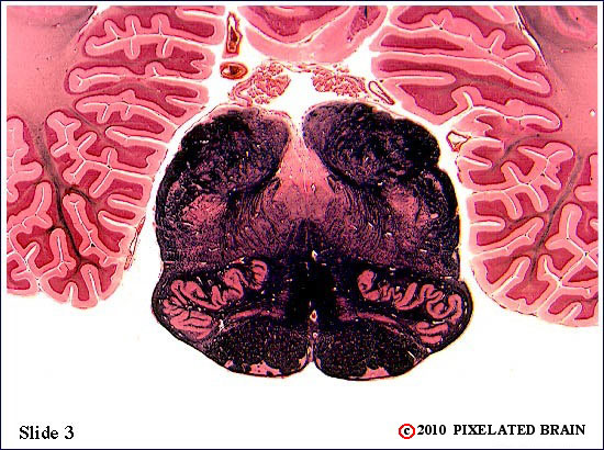
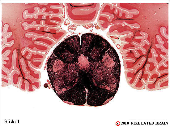
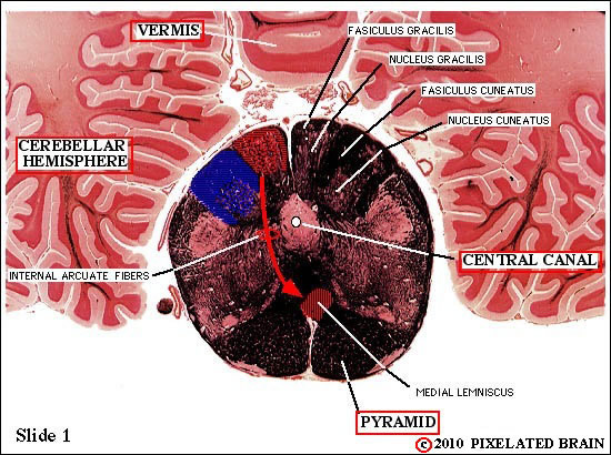
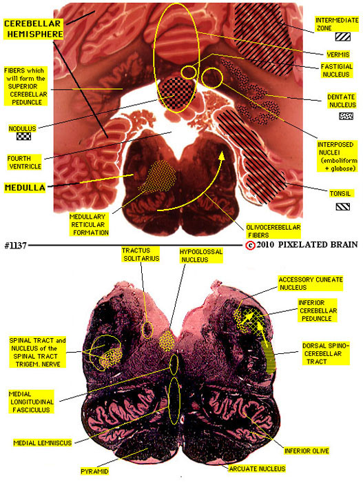
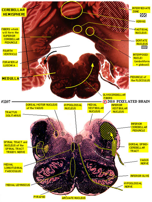
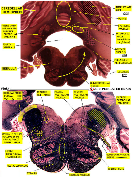
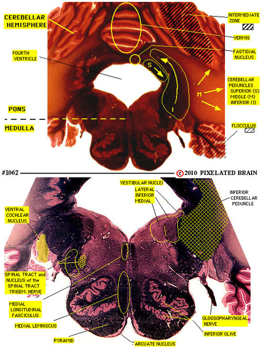
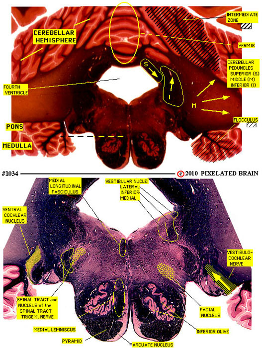
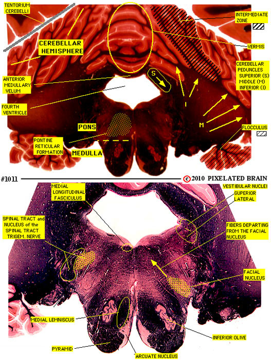
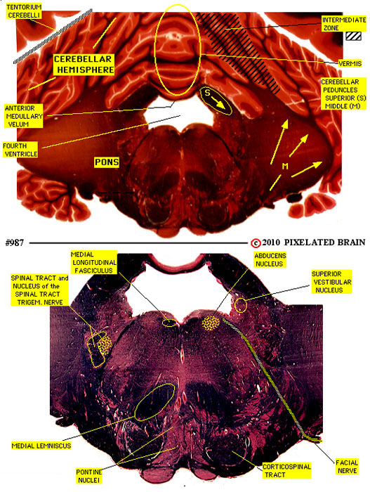
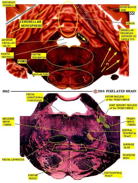
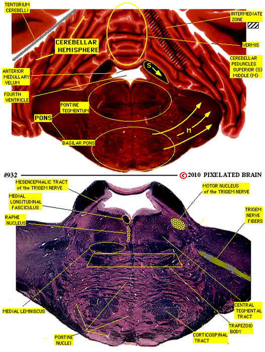
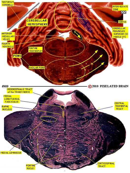
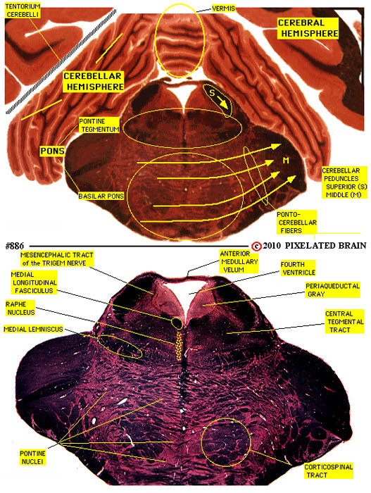
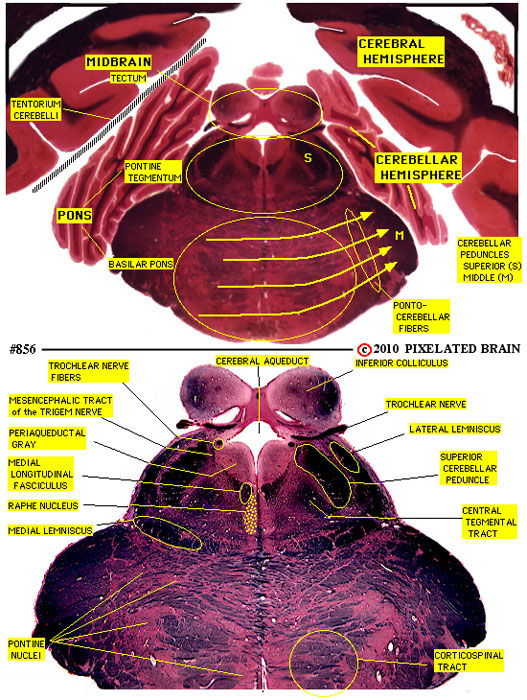
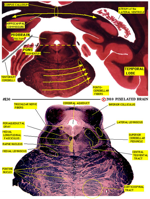
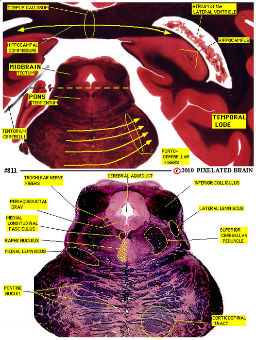
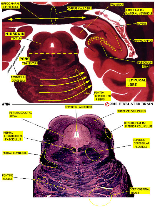
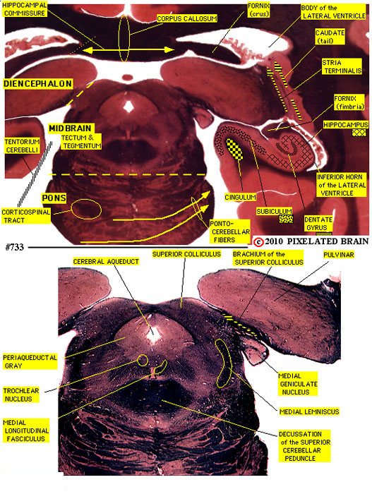
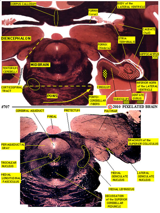
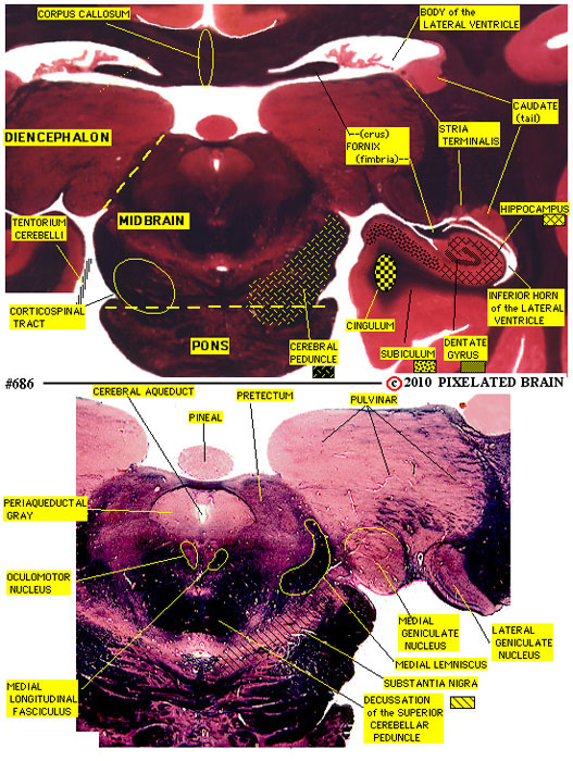
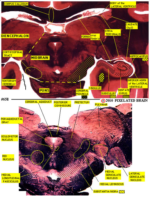
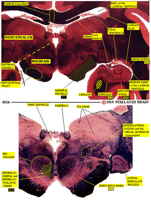
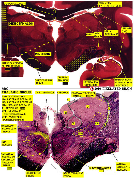
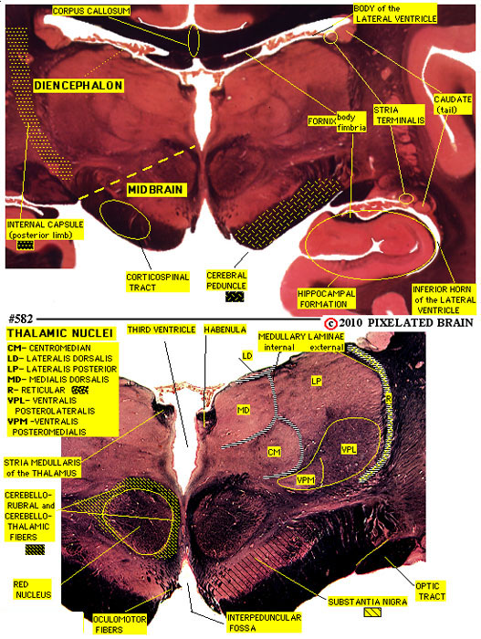
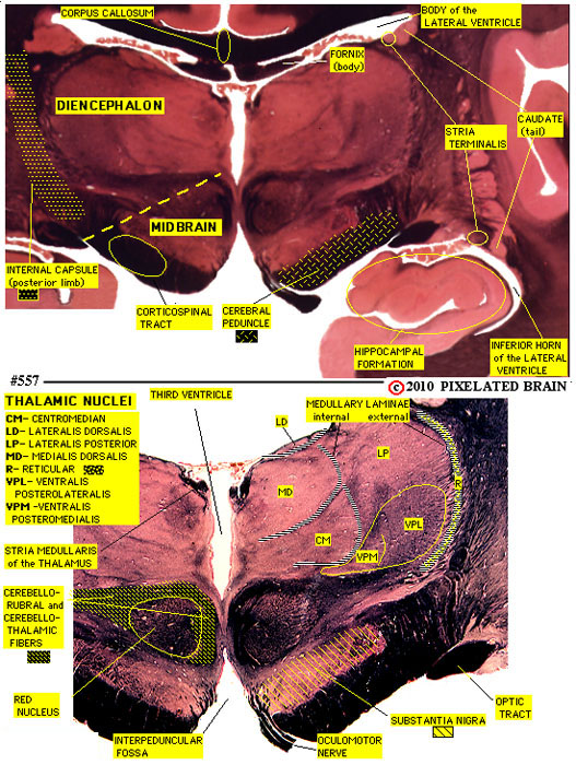
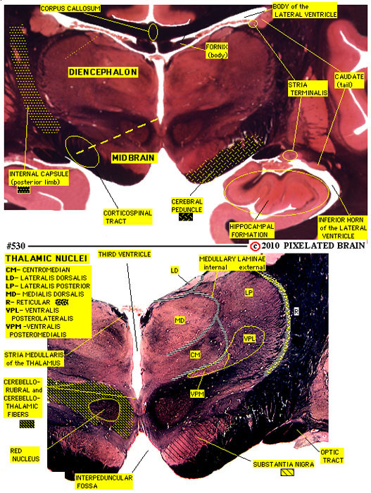
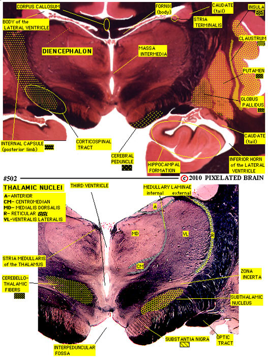
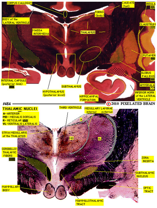
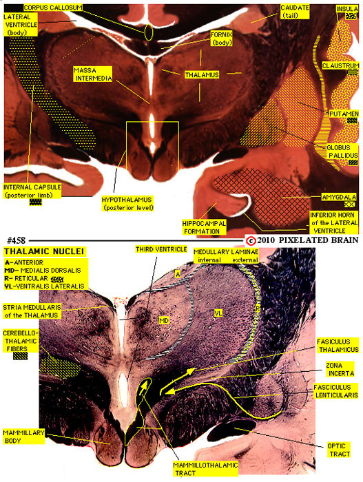
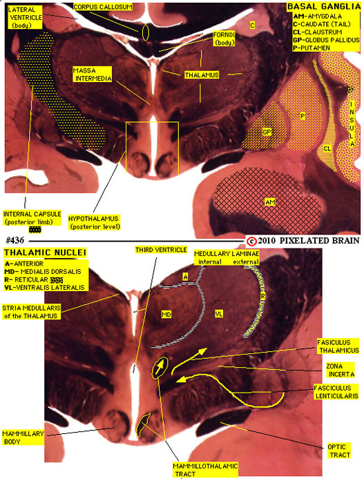
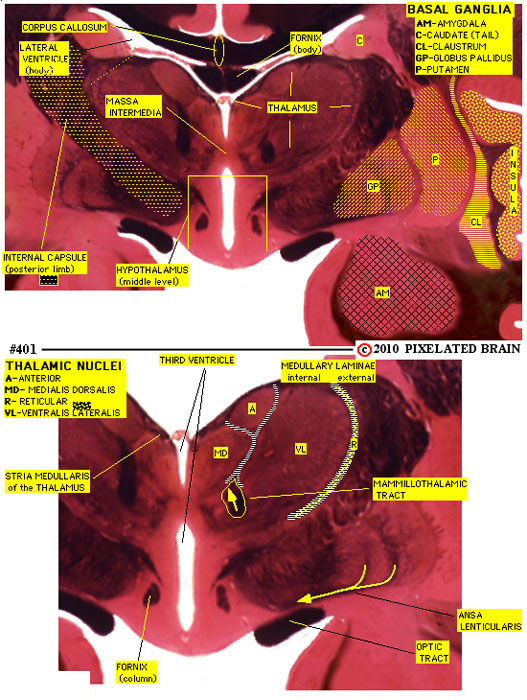
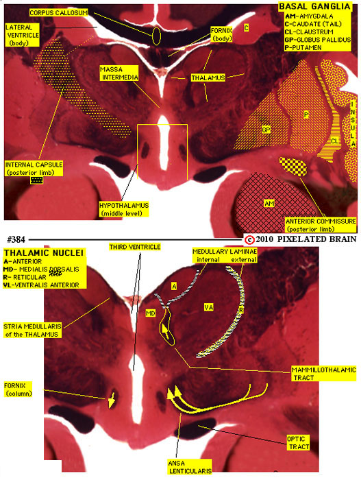
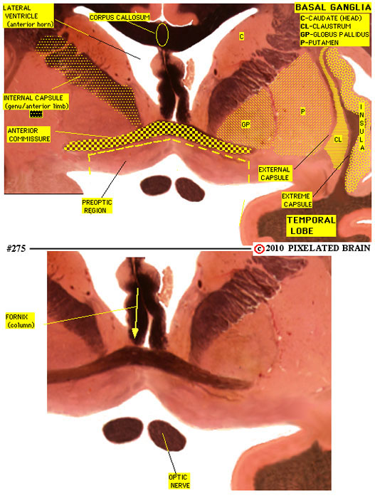
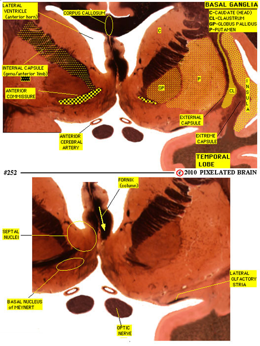
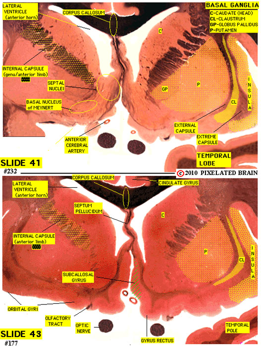
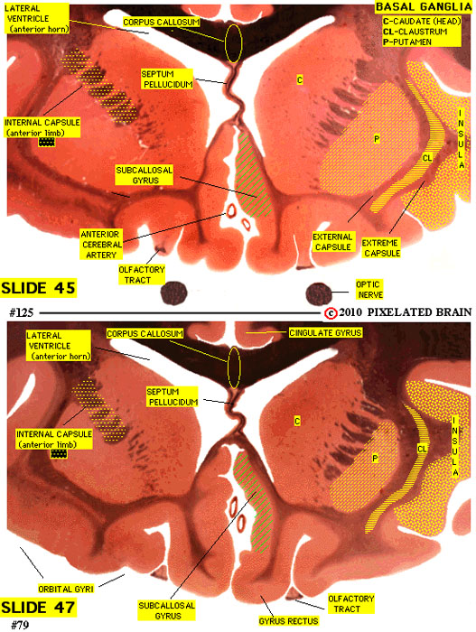
The Pixelated Brain slide series was created several years ago by sectioning a human brain in the frontal (coronal) plane. The specimen was trimmed prior to cutting the serial sections, removing much of the hemispheres but leaving the brainstem, diencephalon, basal ganglia and cerebellum intact. The 15 micron sections were stained using the Weil myelin method (which colors myelinated axons black) and counter stained with carmine to color cellular areas light pink. Slide 3 is a typical slide through the brainstem and overlying cerebellum. There are about 1400 slides in the series and roughly every 30th slide was selected for use in this atlas. The most caudal slide, Slide1, passes through the caudal medulla. The most rostral slide, Slide 47, passes just rostral to the interventricular foramen. Slide 5 is a typical atlas view. As you can see, the upper half of the page contains a low mag version of the slide, showing the tissue which surrouns the brainstem - in this case, the cerebellum. The lower half of the page displays a high mag view of the brainstem. When you view the atlas on a computer, an additional panel contains more information about each section - see the computer version of Slide 5 for an example.
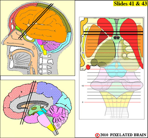
PLANE OF SECTION
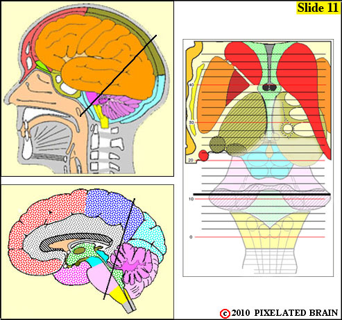
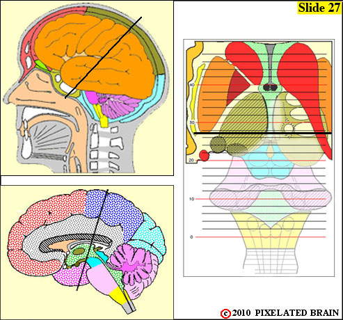
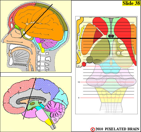
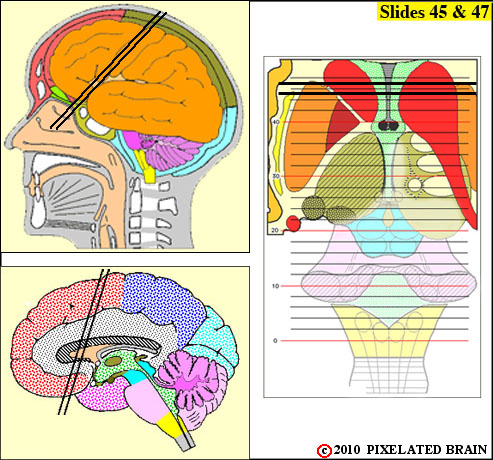
As Figure 2-18 demonstrates, the bend in the neural axis of the brain at the midbrain creates a problem for anyone making serial sections through this complex organ. For example, if the plane of section is set to produce true cross sections through the medulla, the more rostral sections will pass through the diencephalon in an almost horizontal manner. The plane of the section used to prepare this atlas is a compromise . While this distorts the expected appearance of some sections the advantage of using a single set of slides is that a pathway like the pyramidal tract can be traced all the way from the internal capsule to the caudal brainstem.
IDENTIFICATION OF STRUCTURES
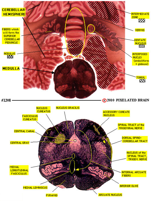
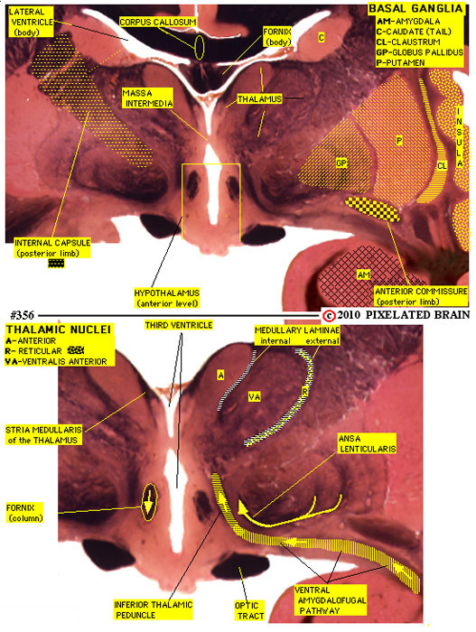
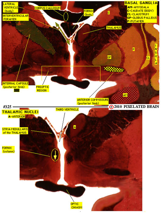
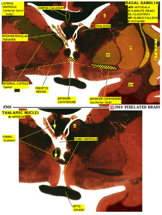
In identifying and labeling structures we have relied heavily on the classic text, An Atlas of the Basal Ganglia, Brain Stem and Spinal Cord by Henry A. Riley, Williams and Wilkins, 1943. A limited number of structures are labeled on the printed version of our atlas, but when using the online version multiple overlays can be called up, each dealing with a specific system or group of nuclei.
HOW TO PRINT YOUR COPY OF THE ATLAS
To view the atlas, scroll down. An alternative is to click on Atlas #2 and call up individual sections. To print a copy, click on Brainstem Atlas for a PDF version. or B or C


Links: DCML

Links: DCML

Links: DCML

Links: DCML

Links: DCML


SITE MAP PIXBRAIN HOME I WANT TO COMMENT