VISUAL PATHWAY - THE OPTIC NERVES
x- PIXBRAIN HOME _ _ MOD 11 HOME _ _ I WANT TO -x
The intracranial course of the optic nerves is quite brief. Obviously, they lie in the space between the basilar surface of the frontal lobe and the dorsal surface of the sphenoid bone, as seen here.
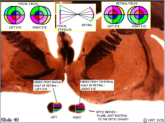
This slide depicts the situation just "in front of" the chiasm. In the upper left we show the color code we will use to describe the visual field. In the center is a little diagram to remind you of what happens (illustrated for the vertical plane only) when the image
is inverted. The diagram on the right shows what this pattern of light would actually look as it reaches the retina. It's easy, however, to confuse retinal maps with visual fields and since information about visual deficits is always expressed in terms visual fields we will omit retinal maps on most views.