PIXBRAIN HOME _ _ MOD 1 HOME _ _ previous _ _ FIGDEV-2-2 _ _ next _ _ I WANT TO
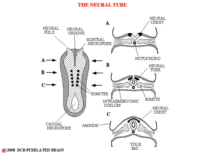
- - On about day 18 following fertilization, the process of neurulation begins. Starting in the cervical region, the neural folds migrate dorsally and fold toward the midline to meet and fuse, forming the first part of the neural tube. Fusion of the folds then extends both rostrally and caudally. For several days the ends of the neural tube remain open, so that the central canal is in communication with the amnionic cavity. The rostral opening, or cranial neuropore, is closed at about day 25 by a membrane called the lamina terminalis. The caudal neuropore closes a few days later.
- - Failure of the cranial neuropore to close leads to anencephaly, a developmental defect which grossly distorts the development of the brain. Failure of the caudal neuropore to close is associated with some form of spina bifida.
- - Some tissue of the neural plate fails to become encorporated in the neural tube and remains just dorsolateral to it. This neural crest tissue ultimately gives rise to many structures, including: 1) the dorsal root (sensory) ganglia of the peripheral nervous system; 2) the ganglia of the autonomic nervous system; 3) the cells of the adrenal medulla and 4) the Schwann Cells that form the myelin that surrounds peripheral nerves.
- - Lateral to the neural tube the mesoderm becomes organized in a segmental fashion into somites which will, ultimately, impose a segmental pattern upon the peripheral nervous system.