LIMBIC SYSTEM - THE ANT HYPOTHALAMUS and AMYGDALA
x- PIXBRAIN HOME _ _ MOD 13 HOME _ _ I WANT TO -x
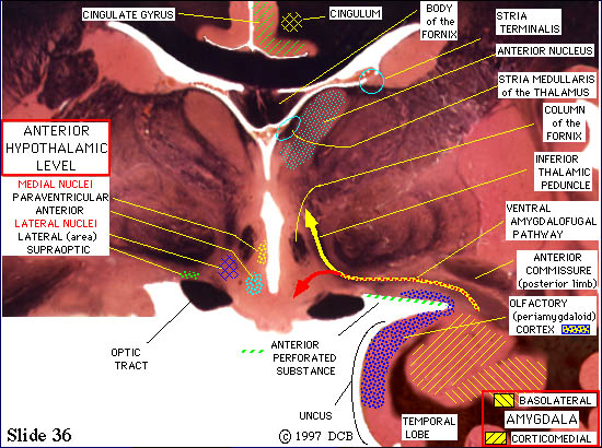
TRIP 1 #6 - Fibers of the lateral olfactory stria continue to terminate in the periamygdaloid cortex and the corticomedial division of the amygdala. From the latter structure olfactory information is relayed to many regions of the forebrain, including the orbitofrontal cortex, the nucleus medialis dorsalis of the thalamus and the basolateral nucleus of the amygdala. Years ago it was beleived that the hippocampus, which lies just caudal to the amygdala in the temporal lobe, received a significant olfactory input, but that does not appear to be the case.
Well, we promised to get you to the amygdala in 5 clicks and here we are. This is the end-point of our trip, but before we go on to trip 2 we want to make a few final points about the amygdala.
1) The amygdala is actually a rather big nucleus. Click on "1 caudal" above a few times and you will see it extends back to slide 32. At that point the nucleus is almost gone, replaced by the rostral tips of the hippocampus and inferior horn of the lateral ventricle. These relationships are shown nicely in Lect. fig 3, Lect. fig 4 and Lect. fig 5. _____(THEN CLICK "1 rostral" ENOUGH TO GET BACK TO THIS SLIDE)
2) You should be aware that the naming of structures in this area is extremely confusing, in large part because they were first identified in more primitive nervous systems. Some anatomists thought the temporal lobe looked "pear shaped" in these brains, thus leading to the term "piriform"; others thought the collateral sulcus, that defines the lateral border of this region (see Fig 2-26)), continued rostrally as the "rhinal sulcus " - hence, the terms entorhinal and perirhinal cortex. In the same vein, there are differences in the way the subnuclei within the nucleus are specified. We show the two major nuclei, but Blumenfeld adds a third - the central nucleus.
3) We have traced just one of the many INPUT PATHWAYS to the amygdala; for a summary of the others, see Lect. fig 15.
4) There are two major OUTPUT PATHWAYS. One of them, the ventral amygdalofugal pathway, is present in this slide. Some of these fibers (the red arrow) terminate in the hypothalamus. Others (the yellow arrow) turn dorsally to end in the Medialis Dorsalis nucleus of the thalamus (to trace it there click "1 caudal" a few times and look for the yellow arrow). The second output pathway, the stria terminalis, takes a more circuitous route. We will follow it when we take trip 2. For a summary of the output pathways, see Lect. fig 16.